A pet-ct scanning imaging method and related imaging method
A PET-CT, scanning imaging technology, applied in the field of medical imaging, can solve the problems of prolonged scanning time and increased radiation dose in irrelevant areas
- Summary
- Abstract
- Description
- Claims
- Application Information
AI Technical Summary
Problems solved by technology
Method used
Image
Examples
Embodiment 1
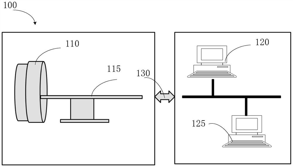
[0064] A PET-CT scanning imaging method is disclosed, comprising the following steps:
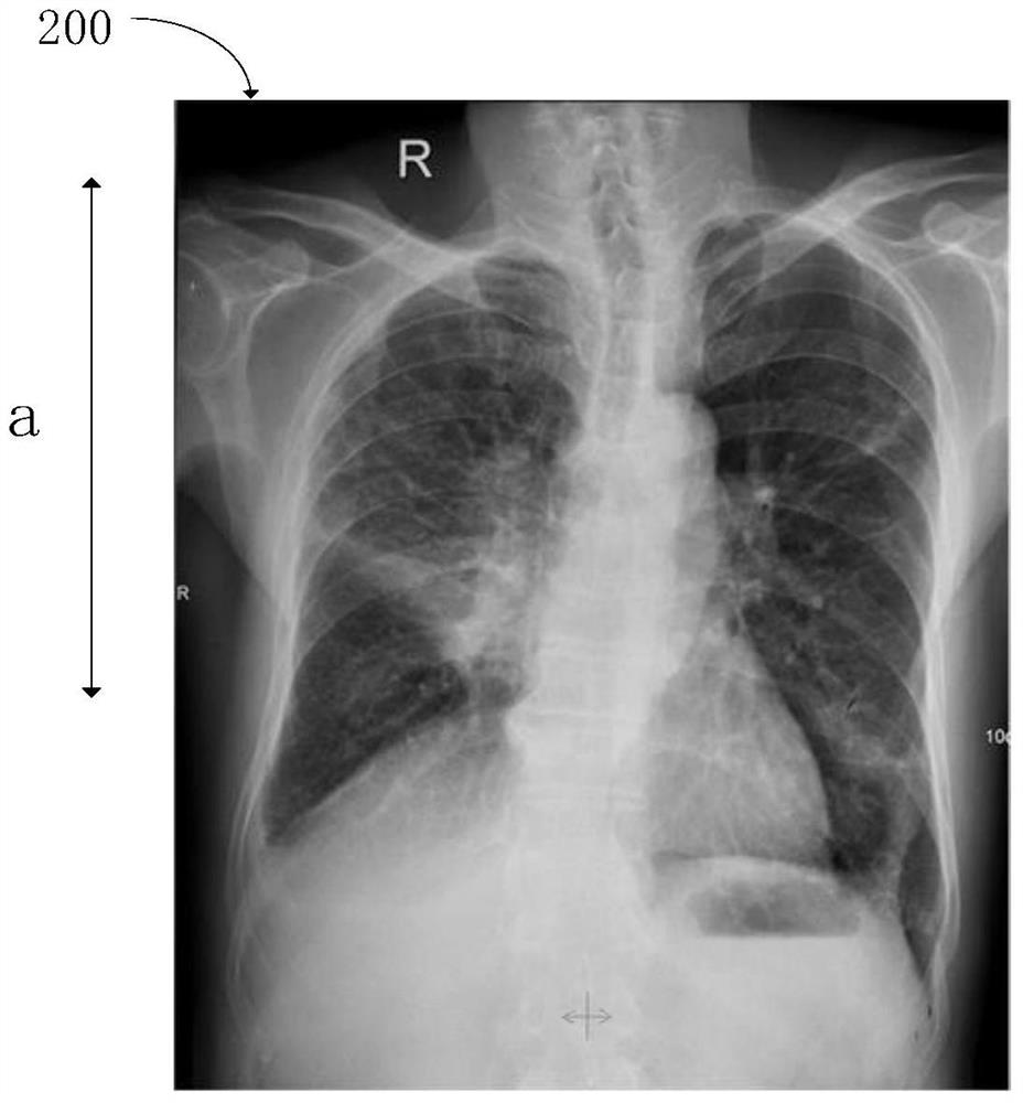
[0065] S101, performing a positioning scan on an area to be imaged, obtaining and displaying a positioning image of the area to be imaged, and the displayed positioning image is used for a user to determine a scanning area;
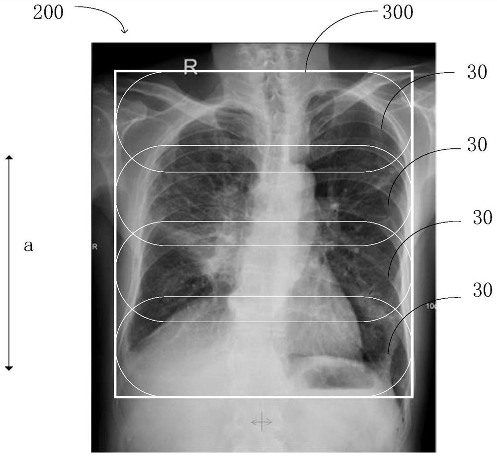
[0066] S102, receiving the scanning area information input by the user, and determining the PET total scanning area according to the scanning area information;
[0067] S103, generating a plurality of PET sub-scanning areas from the PET total scanning area, each PET sub-scanning area corresponds to a PET scanning bed, and each PET sub-scanning area completely covers the PET total scanning area after being superimposed;
[0068] S104, performing a CT scan;
[0069] S105, performing a multi-bed PET scan; and S106, reconstructing a PET-CT image from the scan data.
[0070] Each step is described in detail below:
[0071] First, step S101 is performed to perform a posit...
Embodiment 2
[0096] The present embodiment discloses a PET-MR scanning imaging method, comprising the following steps:
[0097] S201, performing a positioning scan on an area to be imaged, obtaining and displaying a positioning image of the area to be imaged, and the displayed positioning image is used for a user to determine a scanning area;
[0098] S202, receiving the scanning area information input by the user, and determining the PET total scanning area according to the scanning area information;
[0099] S203, generating a plurality of PET sub-scanning areas from the PET total scanning area, each PET sub-scanning area corresponds to a PET scanning bed, and each PET sub-scanning area completely covers the PET total scanning area after being superimposed;
[0100] S204, performing an MR scan;
[0101] S205, performing a multi-bed PET scan;
[0102] S206, reconstruct a PET-MR image from the scan data.
[0103] The implementation process of steps S201 to S206 may refer to the implemen...
Embodiment 3
[0109] The present embodiment discloses a SPECT scanning imaging method, comprising the following steps:
[0110] S301, performing a positioning scan on an area to be imaged, obtaining and displaying a positioning image of the area to be imaged, and the displayed positioning image is used for a user to determine a scanning area;
[0111] S302, receiving the scanning area information input by the user, and determining the SPECT total scanning area according to the scanning area information;
[0112] S303, generating a plurality of SPECT sub-scanning regions from the SPECT total scanning region, each SPECT sub-scanning region corresponds to a SPECT scanning bed, and the SPECT sub-scanning regions are superimposed to completely cover the SPECT total scanning region;
[0113] S304, performing transmission scanning;
[0114] S305, performing a multi-bed SPECT scan; and S306, reconstructing a SPECT image from the scan data.
[0115] The SPECT scanning imaging method is similar to ...
PUM
 Abstract
Description
Claims
Application Information
 - R&D
- Intellectual Property
- Life Sciences
- Materials
- Tech Scout
- Unparalleled Data Quality
- Higher Quality Content
- 60% Fewer Hallucinations
Browse by: Latest US Patents, China's latest patents, Technical Efficacy Thesaurus, Application Domain, Technology Topic, Popular Technical Reports.
© 2025 PatSnap. All rights reserved.Legal|Privacy policy|Modern Slavery Act Transparency Statement|Sitemap|About US| Contact US: help@patsnap.com


