
Fluorescence three-dimensional imaging tumor available receptor density quantitative method based on PET guide irradiation

A radiofluorescence and three-dimensional imaging technology, applied in the field of medical imaging, can solve the problems of complex experimental operation, quantification, different emission bands, etc., to simplify complexity, ensure accuracy, and reduce radioactive pollution.

Inactive Publication Date: 2015-09-30
XIDIAN UNIV
View PDF2 Cites 5 Cited by
  • Summary
  • Abstract
  • Description
  • Claims
  • Application Information

AI Technical Summary

Problems solved by technology

This technology has the advantage of no radioactive pollution, but it requires dual probes with close molecular weight, similar structure, the same spectral excitation band but different emission bands for subsequent spectral signal separation, etc. The requirements are too harsh, so the experimental operation process of this method is extremely complex, and the method cannot simultaneously quantify tumor metabolic information
[0006] In summary, for the quantification of the available receptor density information of tumors, the existing technologies and methods cannot realize the non-invasive in vivo quantification of receptor density in the tumor area, and at the same time cannot avoid excessive radioactive contamination and complex experimental design. defect

Method used


Image

  • Fluorescence three-dimensional imaging tumor available receptor density quantitative method based on PET guide irradiation
  • Fluorescence three-dimensional imaging tumor available receptor density quantitative method based on PET guide irradiation
  • Fluorescence three-dimensional imaging tumor available receptor density quantitative method based on PET guide irradiation

Examples


Embodiment Construction

[0062] The present invention will be further described below in conjunction with the accompanying drawings. It should be noted that this embodiment is based on the technical solution, and provides detailed implementation and specific operation process, but the protection scope of the present invention is not limited to the present invention. Example.

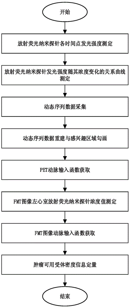
[0063] Such as figure 1 As shown, the method for quantifying available receptor density in tumors based on PET-guided radiofluorescence three-dimensional imaging includes the following steps:

[0064] (1) Measurement of Luminescence Intensity of Radiofluorescent Nanoprobes at Each Time Point

[0065] From 0nmol to 3nmol as the upper limit, 31 radiofluorescence nanoprobes Gd2O2S:Tb-GX1 concentration gradients were set at 0.1nmol concentration intervals, and they were uniformly mixed with 100uci of PET probe 18F-FDG and placed in the optical imaging chamber immediately. Continuous observation, according to the dynamic time frame...


PUM


Abstract

The invention discloses a fluorescence three-dimensional imaging tumor available receptor density quantitative method based on PET guide irradiation. Based on the phenomenon that rare earth doping objects are stimulated by high-energy rays emitted by a radioactive nuclide probe to emit fluorescence, a specific radiation nanometer molecular probe is constructed, fluorescence image information emitted by a target molecular probe is further used, PET image information of the nuclide probe serves as reference input for fluorescence three-dimensional imaging quantitative analysis, and therefore the aim of quantifying tumor available receptor density information is achieved. The method solves the problems that through an existing technology and an existing method, non-intrusive type living body quantifying on tumor area receptor density cannot be achieved, and meanwhile too-large radioactive pollution, complex experimental design and the like are avoided. The method has the advantages that non-intrusive type living body quantifying on the tumor area receptor density and metabolism information can be achieved at the same time, the radioactive pollution is small, and experiment operation is simple; the method can be applied to the field of medical optical imaging.

Description

technical field [0001] The invention relates to the field of medical imaging, in particular to a method for quantifying available tumor receptor density based on PET-guided radiofluorescence three-dimensional imaging. This method can be applied to small animal experiments and pre-clinical experiments such as tumor detection, new drug development, and efficacy evaluation to obtain density information of targeted receptors. Background technique [0002] Tumor occurrence is a multi-step process in which cell genome changes or its integrity is destroyed, resulting in the loss or loss of control of normal physiological functions of cells and malignant transformation of cells, also known as cell carcinogenesis. Most tumor cells mainly exhibit the following physiological characteristics in the process of carcinogenesis: autonomous production of growth factors, insensitivity to growth inhibitory signals, resistance to programmed cell death, infinite continuous division and prolifera...

Claims


Application Information

Patent Timeline
Patent Type & Authority Applications(China)
IPC IPC(8): A61B6/00
Inventor 陈雪利代云鹏梁继民殷继鹏曹旭詹勇华田捷
Owner XIDIAN UNIV
Learn More