
Automatic knee cartilage image partitioning method based on SVM (support vector machine) and elastic region growth

An elastic region, automatic segmentation technology, applied in the field of image processing, can solve the problems of subdivision, unsatisfactory segmentation results, over-segmentation, etc.

Inactive Publication Date: 2015-07-29
CHONGQING UNIV
View PDF3 Cites 28 Cited by
  • Summary
  • Abstract
  • Description
  • Claims
  • Application Information

AI Technical Summary

Problems solved by technology

However, these algorithm studies also have some limitations: first, the algorithm needs to initialize the contour and the requirements are very high, and the cartilage of different shapes in each sequence image needs to roughly represent the target contour, otherwise it will lead to premature convergence and unsatisfactory segmentation results
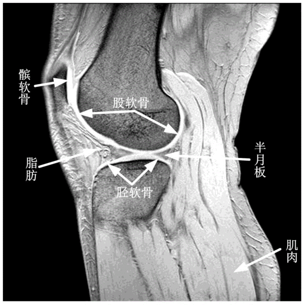
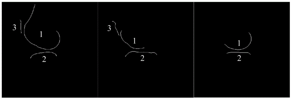
[0005] 1. Due to the complexity of the texture and shape of the knee joint image, it is interfered by many non-cartilage edges, and there are many false edges in the edges detected by the traditional edge detection method. Even if some scholars propose to use SVM for edge classification, it is often because of the selected The feature parameters are limited, only the cartilage edge and non-cartilage edge can be identified, and no further subdivision is made for femur-cartilage, tibia-cartilage and patella-cartilage
[0006] 2. The traditional region growing method compares with similar pixel values ​​in the field according to the similarity criterion. The threshold value is usually set at a fixed value. Due to the difference in gray level between different sequences of images of different people, it will lead to over-segmentation or undersegmentation

Method used


Image

  • Automatic knee cartilage image partitioning method based on SVM (support vector machine) and elastic region growth
  • Automatic knee cartilage image partitioning method based on SVM (support vector machine) and elastic region growth
  • Automatic knee cartilage image partitioning method based on SVM (support vector machine) and elastic region growth

Examples


Embodiment Construction

[0052] The present invention will be further described below in combination with specific embodiments and accompanying drawings. The specific embodiments described here are only used to explain the present invention, not to limit the present invention.

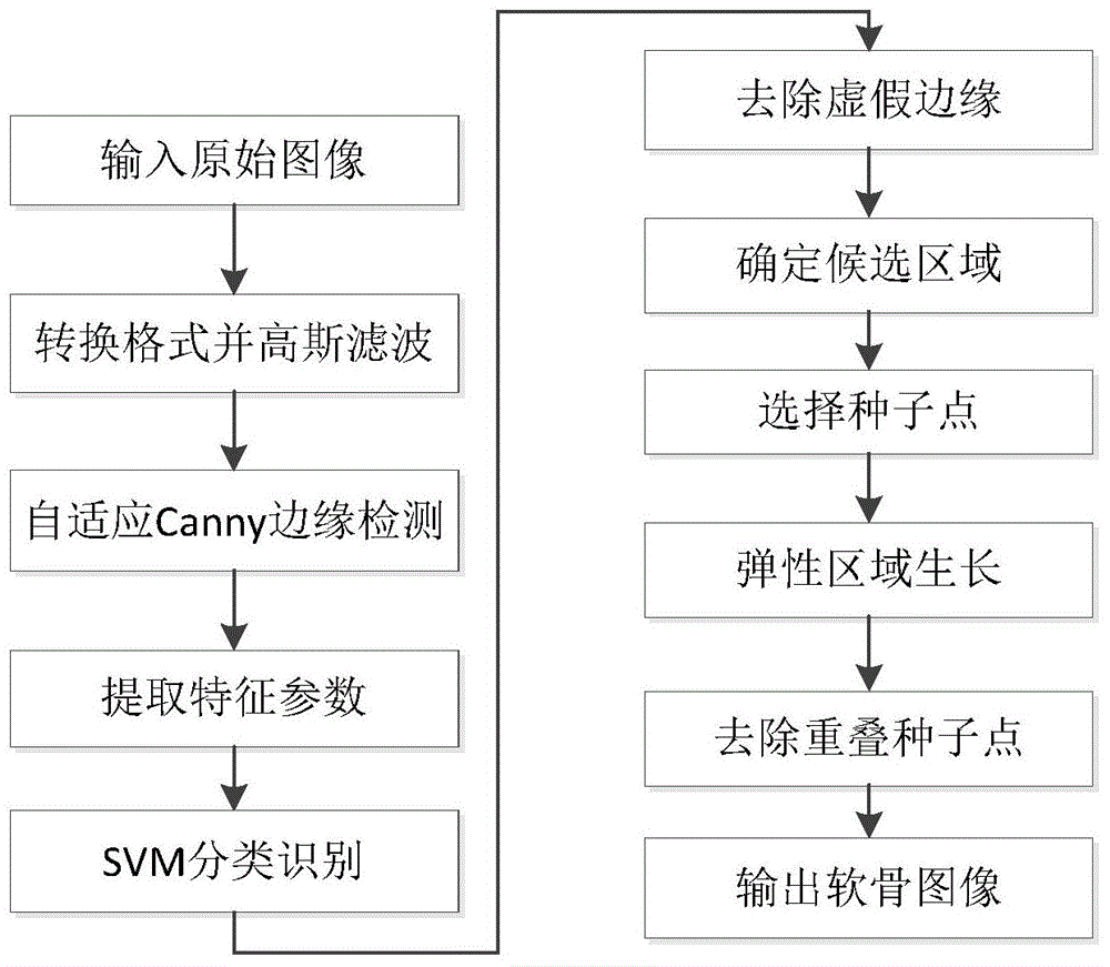
[0053] Such as figure 1 As shown, an automatic segmentation method of knee cartilage images based on SVM and elastic region growth is carried out according to the following steps:

[0054] Step 1: convert the MRI image of the knee joint into a grayscale image and perform Gaussian filtering;

[0055] The sagittal MRI image of the right knee joint of 5 healthy adult males (age range 20-25 years old) without joint medical history used in this embodiment is used as the research object, and the image is obtained using a 1.5T Siemens scanner, using a T2-weighted fat-suppressed sequence ( Sagittal slice thickness: 2.5mm, FOV: 160×160mm, resolution 384×384, TR: 1363ms, TE: 4.42ms, deflection angle: 60°, number of slices: about 25). ...


PUM


Abstract

The invention discloses an automatic knee cartilage image partitioning method based on SVM (support vector machine) and elastic region growth. The method comprises the steps of firstly extracting main image edges by use of a self-adaption Canny edge detection algorithm, extracting multiple image characteristics of the edges, classifying the edges in combination with an SVM algorithm, finishing cartilage edge positioning, performing seed point and cartilage pixel region selection on the basis of the cartilage edges, then performing initiative cartilage partitioning based on selection results by use of elastic region growth, and finally obtaining the knee cartilage partitioning result. Experimental results indicate that the method can accurately, rapidly and automatically partition different knee cartilages in knee joint MRI, wherein the average DSCs of femora cartilage, shank cartilage and patella cartilage are respectively up to 0.8543, 0.8280 and 0.8703; the result has high consistency compared with the manual partitioning result; the defects that excessive partitioning or inaccurate partitioning and the like in the results of traditional partitioning methods are effectively overcome.

Description

technical field [0001] The invention relates to image processing technology, in particular to a knee cartilage image automatic segmentation method based on SVM (Support Vector Machine, support vector machine) and elastic region growth. Background technique [0002] The knee joint is the joint with the most complex structure and the most vulnerable joints in the human body. Its common diseases include arthritis, bone tumors, etc., and these diseases are often accompanied by the degeneration, destruction and morphological changes of articular cartilage. Early diagnosis is very important. As a non-invasive examination method, magnetic resonance imaging has become the main means to evaluate the morphology and function of cartilage. Segmenting articular cartilage by MRI imaging and then calculating its thickness, volume and other parameters can realize quantitative evaluation of cartilage and provide a strong diagnostic basis for clinical medicine, so as to take early preventive...

Claims


Application Information

Patent Timeline
IPC IPC(8): G06T7/00G06K9/62
CPCG06T2207/30008G06F18/2411
Inventor 王品何璇李勇明李帆吴烨
Owner CHONGQING UNIV
Learn More