Eureka AIR delivers breakthrough ideas for toughest innovation challenges, trusted by R&D personnel around the world.

Method for cell imaging by adopting polarized light microscope to observe nano particles

A polarizing microscope and nanoparticle technology, applied in the field of medical detection, to achieve the effect of strong background reduction

Inactive Publication Date: 2015-06-17
李乐 +2
View PDF1 Cites 9 Cited by
  • Summary
  • Abstract
  • Description
  • Claims
  • Application Information

AI Technical Summary

Problems solved by technology

To date, common polarizing microscopes have not been used experimentally to observe individual nanoparticles, mainly because: on the one hand, there is less and less awareness of materials analysis using polarizing microscopy due to the existence of other microscopic imaging techniques; on the other hand, , the nanoparticles are all in the nanometer scale, because the size is too small, if the nanomaterials do not have very strong optical properties, it is difficult to be observed

Method used


Image

  • Method for cell imaging by adopting polarized light microscope to observe nano particles
  • Method for cell imaging by adopting polarized light microscope to observe nano particles
  • Method for cell imaging by adopting polarized light microscope to observe nano particles

Examples


Embodiment

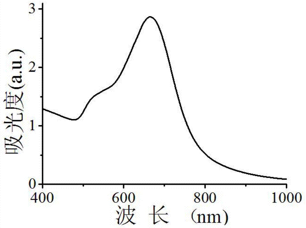
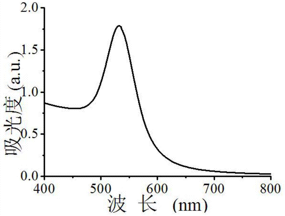
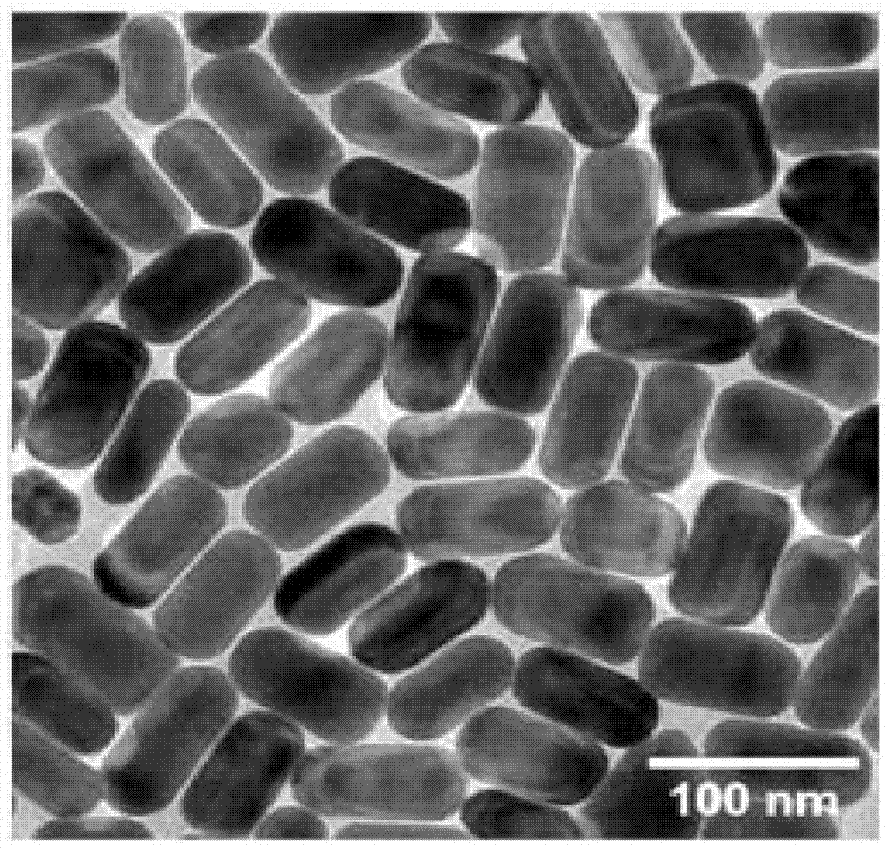
[0026] 1. Reagents

[0027] Sodium citrate, chloroauric acid (99.99%, HAuCl 4 3H2O), cetyltrimethylammonium bromide (99%, CTAB) were all purchased from Shanghai Sinopharm Company; sodium borohydride (98%, NaBH 4 ), ascorbic acid (99.7%, AA), silver nitrate (99.8%, AgNO 3 ), mercaptoundecyltrimethylammonium bromide (99%, MUTAB) were purchased from Sigma-Aldrich Company. The above reagents were prepared with ultrapure water when used.

[0028] Aqua regia (volume ratio HCl:HNO 3 =3:1) Perform soaking and cleaning.

[0029] 2. Synthesis of antibody-modified gold nanorods

[0030] 1) Synthesis of gold nanoparticle seeds: In a serum bottle, inject 48 μL of 0.1M ice-cold sodium borohydride containing 0.1M CTAB and 0.00025M HAuCl 4 In the 4mL solution, the solution needs to stand in the solution at 28°C for more than 2 hours before use.

[0031] 2) In 20mL 0.1M CTAB solution, add 426μL 24.28mM HAuCl in sequence 4 ,0.5ml 4mM AgNO 3 and 105μL 0.1M AA; when the solution turns f...


PUM


Abstract

The invention discloses a method for cell imaging by adopting a polarized light microscope to observe nano particles; a single gold nanorod is observed by adopting the polarized light microscope; the polarized light microscope is found to be capable of selective imaging of an anisotropic nano material; under the polarized light microscope, scattering light spots of the gold nanorod has smaller size, the background value of imaging can be significantly reduced, and accordingly, the signal-to-noise ratio of an image is improved by at least 3 times or more. Due to the unique optical properties, the anisotropic nano material and the polarized light microscope have great application potentiality in imaging and detection of cancer cells.

Description

technical field [0001] The invention relates to the technical field of medical detection, in particular to a method for observing nanoparticles with a polarizing microscope for cell imaging. Background technique [0002] In recent years, gold nanorods (GNRs) have aroused in-depth research interests of scientists and have been widely promoted and applied. In the field of imaging, many optical properties of gold nanorods have been exploited to develop in vivo and in vitro imaging techniques. For example, gold nanosensors have very high optical stability and high optical absorption and scattering efficiencies due to the surface plasmon resonance effect. In fact, they have higher light absorption and scattering cross-sectional areas than other shapes of nanorod materials. The long-axis plasmon resonance absorption peak of gold nanorods can be easily adjusted from the visible region to the near-infrared region by changing the aspect ratio (ratio of length and diameter), and bio...

Claims


Application Information

Patent Timeline
IPC IPC(8): C12Q1/02
Inventor 李乐金鑫贺志红
Owner 李乐
Why Eureka
Eureka Blog
Learn More