Method for separating cells from blood and cultivating the cells and method for cloning non-human animal
A technology for culturing cells and neutrophils, which is applied in the field of cloning non-human animals, can solve the problems such as the need to improve the source of nuclear donor cells, and achieve the effects of improving animal welfare, increasing cell survival rate, and simple collection operation
- Summary
- Abstract
- Description
- Claims
- Application Information
AI Technical Summary
Problems solved by technology
Method used
Image
Examples
Embodiment 1
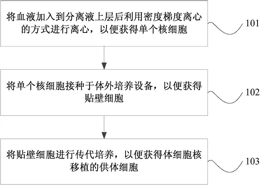
[0067] Example 1 Obtaining mononuclear cells from blood
[0068] Add 125IU heparin to the 15mL conical centrifuge tube in advance, and make it evenly distributed on the tube wall of the conical centrifuge tube by shaking. About 12 mL of the peripheral blood of the miniature pig was collected by directly drawing blood from the porcine anterior vena cava with a 50 mL syringe into the conical centrifuge tube added with 15 mL of heparin. The conical centrifuge tube loaded with peripheral blood was transported to the laboratory in a 4°C ice box.
[0069] Take another 15mL conical centrifuge tube, add 5mL of neutrophil separation solution (containing 57g / L polysucrose and 90g / L sodium diatrizoate) to each of them, and carefully pipette the 15mL Slowly add 5mL of the collected anticoagulated peripheral blood to the wall of the conical centrifuge tube close to the surface of the neutrophil separation medium, and ensure that the anticoagulant peripheral blood does not mix with the neu...
Embodiment 2
[0071] Embodiment 2 Mononuclear cells cultured in vitro
[0072] Add 2 mL of low sugar-MEM, 15% FBS, [1×] non-essential amino acids, 10 ng / mL bFGF and 30 IU / mL of the first adherent cell culture medium of heparin, and use a Pasteur pipette to evenly disperse the mononuclear cell pellet in the first adherent cell culture medium. The first adherent cell culture medium containing mononuclear cells was transferred to a gelatin-coated 35 mm culture dish, and then placed in a carbon dioxide incubator at 37 degrees Celsius for cultivation.
[0073] After 48 hours of culturing, the 35mm culture dish loaded with mononuclear cells was taken out of the incubator, and after discarding the culture medium of the first adherent cells, PBS was added for rinsing. After discarding the PBS, add a second adherent cell medium containing MEM-α, 15% FBS, [1×] non-essential amino acids, and 10 ng / mL bFGF to the 35 mm culture dish loaded with mononuclear cells, and culture The dish was returned to ...
Embodiment 3
[0074] Subculture of embodiment 3 mononuclear cells
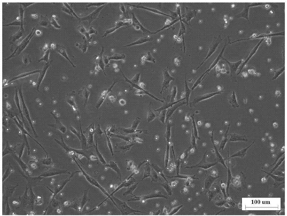
[0075] When the adherent mononuclear cells are observed under the microscope as clumps of contact inhibitors (see Figure 4 ), discard the second culture medium for adherent cells in the culture dish; then add PBS buffer to wash the cells twice; after discarding the PBS buffer, add 200 μL of 0.05 % trypsin-200mg / L EDTA, and place the cells in a carbon dioxide incubator at 37 degrees Celsius for digestion. When observed under the microscope, the adherent cells gradually become round and bright, and gradually break away from the culture dish and float in the trypsin. , add 1mL of the second adherent cell culture medium to the culture dish, and pipette through a Pasteur pipette to mix the mononuclear cells with the added fresh second adherent cell culture medium evenly, so as to obtain a cell suspension; The cell suspension was transferred to a gelatin-coated culture device for subculture. The adherent mononuclear cells that...
PUM
 Abstract
Description
Claims
Application Information
 - R&D
- Intellectual Property
- Life Sciences
- Materials
- Tech Scout
- Unparalleled Data Quality
- Higher Quality Content
- 60% Fewer Hallucinations
Browse by: Latest US Patents, China's latest patents, Technical Efficacy Thesaurus, Application Domain, Technology Topic, Popular Technical Reports.
© 2025 PatSnap. All rights reserved.Legal|Privacy policy|Modern Slavery Act Transparency Statement|Sitemap|About US| Contact US: help@patsnap.com


