
Image stitching method and device

An image stitching and image technology, applied in the field of medical image processing, can solve the problems of low image registration accuracy and long stitching processing time, and achieve the effect of improving time performance, reducing the probability of easily falling into local optimal values, and the method is simple

Active Publication Date: 2015-01-07
SHANGHAI UNITED IMAGING HEALTHCARE
View PDF5 Cites 31 Cited by
  • Summary
  • Abstract
  • Description
  • Claims
  • Application Information

AI Technical Summary

Problems solved by technology

[0008] The problem solved by the invention is that the image registration accuracy is low in the process of image stitching, and the stitching process takes a long time

Method used


Image

  • Image stitching method and device
  • Image stitching method and device
  • Image stitching method and device

Examples


Embodiment Construction

[0048] In the prior art, in the angiographic examination, MRA scans cannot obtain images of a large field of view at one time, and several consecutive scans of the target large field of view are required. Two adjacent scans include overlapping areas, and a series of images containing overlapping Multi-segment subtraction angiography volume data of the region, and then need to register a series of segment subtraction angiography volume data, and finally stitch multiple segment subtraction angiography volume data into a panoramic subtraction angiography volume data Shadow angiography volume data.

[0049] However, in the registration process of subtraction angiography volume data stitching, it is difficult to obtain better registration due to factors such as low signal-to-noise ratio of subtraction angiography images and large differences in image information in overlapping areas of image stitching. As a result, the accuracy of the image registration algorithm is low, and due to...


PUM


Abstract

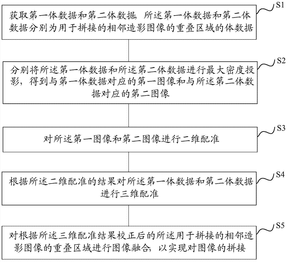
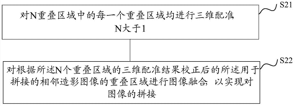
The invention provides an image stitching method and device. The image stitching method and device are used for stitching three-dimensional digital subtraction angiography images. The image stitching method comprises the steps of obtaining first volume data and second volume data, wherein the first volume data and the second volume data are volume data of overlapped areas, used for stitching, of every two adjacent angiography images; maximum intensity projection is conducted on both the first volume data and the second volume data, so that a first image corresponding to the first volume data and a second image corresponding to the second volume data are obtained; two-dimensional registration is conducted on the first image and the second image; according to a registration result, three-dimensional registration is conducted on the first volume data and the second volume data; image fusion is conducted on the overlapped areas, used for stitching, of every two adjacent angiography images after correction is conducted according to a three-dimensional registration result, and therefore stitching of the images is achieved. By the adoption of the image stitching method, the accuracy of a three-dimensional registration algorithm can be effectively improved, and the stitching time of the images can be effectively shortened.

Description

technical field [0001] The invention relates to a medical image processing method, in particular to an image splicing method and an image splicing device. Background technique [0002] Angiography is currently the main method for diagnosing vascular diseases, and it is an interventional detection method. Angiography refers to injecting a contrast agent into a blood vessel, and scanning with an imaging device to visualize the target blood vessel, so that the stenosis and occlusion of the blood vessel can be found. Usually, the angiography refers to digital subtraction angiography. Specifically, before injecting the contrast agent, the first imaging is performed first, and the image is converted into a digital signal by a computer for storage. After the contrast agent is injected, the imaging is performed again and converted into Digital signal; two digital signals are subtracted to obtain a subtraction image, which eliminates the influence of bone and soft tissue, so that bl...

Claims


Application Information

Patent Timeline
IPC IPC(8): G06T5/50G06T7/00
Inventor 于文君
Owner SHANGHAI UNITED IMAGING HEALTHCARE
Learn More