Eureka AIR delivers breakthrough ideas for toughest innovation challenges, trusted by R&D personnel around the world.

Method for separating and culturing human epidermal stem cells

An epidermal stem cell and culture method technology, applied in the field of separation and culture of human epidermal stem cells, can solve the problems of limited cell expansion, complex methods, difficult conditions, etc. Good results

Inactive Publication Date: 2014-06-18
THE FIRST AFFILIATED HOSPITAL OF THIRD MILITARY MEDICAL UNIVERSITY OF PLA
View PDF0 Cites 14 Cited by
  • Summary
  • Abstract
  • Description
  • Claims
  • Application Information

AI Technical Summary

Problems solved by technology

However, the in vitro culture of epidermal stem cells has been delayed in clinical application.
The traditional human epidermal stem cell culture method needs to be co-cultured with 3T3 trophoblast cells and needs to be added with serum. The method is complicated, the conditions are difficult to be consistent, the stability is poor, and the cell differentiation is fast. Generally, the number of cells can only be transferred to the 3rd to 5th generation. Affected by trophoblasts, it cannot be applied clinically

Method used


Image

  • Method for separating and culturing human epidermal stem cells
  • Method for separating and culturing human epidermal stem cells
  • Method for separating and culturing human epidermal stem cells

Examples


Embodiment 1

[0043] Embodiment 1 adopts the method of the present invention to cultivate human epidermal stem cells

[0044] (1) Soak the removed human foreskin tissue in 0.5% povidone iodine for one minute, rinse thoroughly with PBS twice until there is no povidone iodine staining with the naked eye. Foreskin tissues were obtained from patients aged 12 to 20 years old who underwent circumcision without urinary tract infection and other diseases in the Department of Urology, Southwest Hospital of Third Military Medical University (informed consent of patients);

[0045](2) Under aseptic conditions, the subcutaneous fat tissue was trimmed as much as possible, and the tissue was trimmed into a piece of skin about 0.5 cm×1 cm in size.

[0046] (3) Add 0.25% dispase II solution to fully cover the tissue block, and digest at 4°C for 12-16 hours; the configuration and use of 0.25% dispase II solution is as follows: dissolve 0.5g Dispase II (Roche) into 100mL 0.1MPBS, 4 ℃, overnight, then stir t...

Embodiment 2

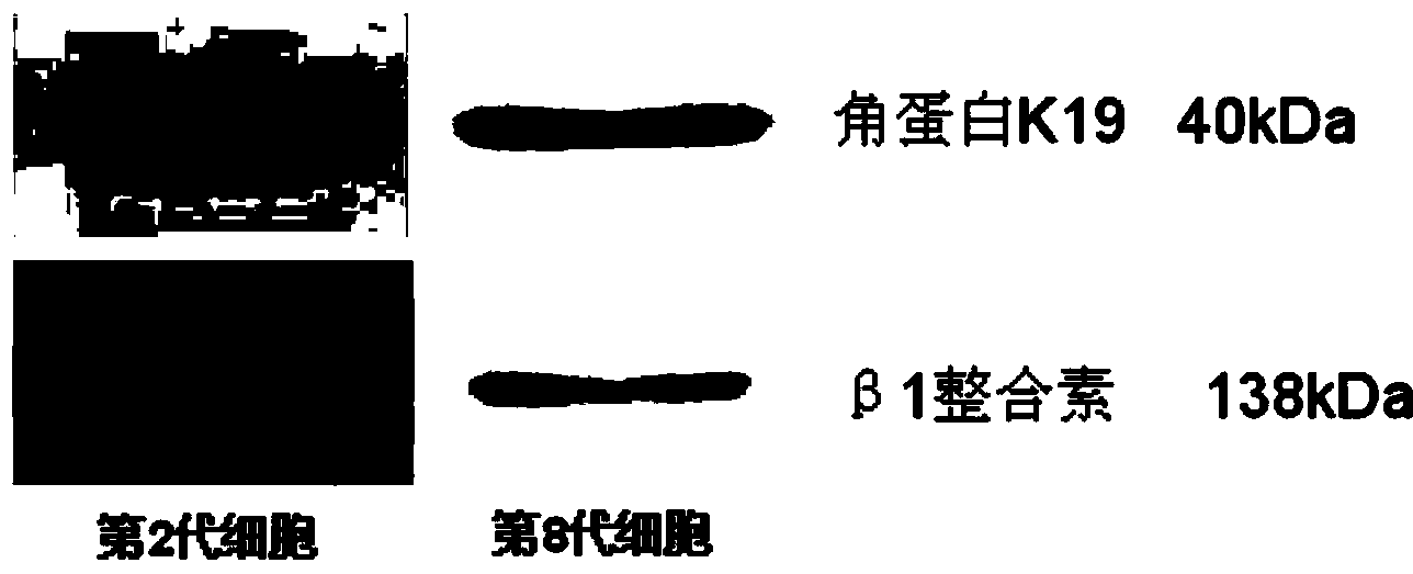
[0063] Example 2 Identification of Primary Cultured Human Epidermal Stem Cells

[0064] (1) WB detection of expression of epidermal stem cell markers β1 integrin and ck19 protein in isolated cultured cells.

[0065] ① Extraction and quantification of protein: at a concentration of 10 5 Inoculate a 6-well plate with cells / mL, inoculate 1 mL in each well, and extract the total protein after the cell density reaches 80%. The specific operation steps are as follows:

[0066] 1) After discarding the medium, wash the cells twice with 0.1MPBS, suck up the PBS with a sample gun, add 200μL of 0.25% trypsin to each well, digest for about 5 minutes, add 1mL of PBS to each well, pipette, and pipette Transfer the cell suspension to a centrifuge tube at 800 rpm, discard the supernatant after 10 minutes, add 250 μL of RIPA and 5 μL of PMSF to each tube, and place it on ice for 10 minutes for digestion.

[0067] 2) Transfer all the cell suspension to a 1.5mLEP tube, and lyse the cells by so...


PUM


Abstract

The invention belongs to the technical field of pathology, and in particular relates to a method for separating and culturing human epidermal stem cells. The technical problem to be solved by the method disclosed by the invention is to provide a new choice for separating and culturing the human epidermal stem cells. The technical scheme is that the method for separating and culturing the human epidermal stem cells comprises the steps of a, sterilizing; b, separating epidermis and dermis; c, collecting; d, culturing; e, sub-culturing. By adopting the method disclosed by the invention, the human epidermal stem cells at least can be sub-cultured to the eighth generation, so that the cell proliferation multiple is greatly increased.

Description

technical field [0001] The invention belongs to the technical field of pathology, and in particular relates to a method for separating and culturing human epidermal stem cells. Background technique [0002] Skin is the largest tissue and organ of the human body. It covers the surface of the human body and has important functions such as resisting microbial invasion, preventing ultraviolet radiation and water loss, regulating body temperature, and maintaining appearance. It is the main content of burn and trauma surgery research. The self-renewal of normal skin and the repair of damaged skin are the result of the joint action of various types of cells, and are the basic requirements for maintaining normal tissue structure and the stability of the internal environment of cells. In terms of burns and trauma, epidermal stem cells have unquestionable potential. They are skin tissue-specific stem cells that play an important role in maintaining epidermal self-renewal, maintaining ...

Claims


Application Information

Patent Timeline
IPC IPC(8): C12N5/074
Inventor 詹日兴罗高兴吴军
Owner THE FIRST AFFILIATED HOSPITAL OF THIRD MILITARY MEDICAL UNIVERSITY OF PLA
Why Eureka
Eureka Blog
Learn More