
Multi-dimensional electrocardiosignal imaging system and method

An imaging method, a technique for electrocardiographic signals, used in medical science, sensors, diagnostic recording/measurement, etc.

Active Publication Date: 2014-06-04
河南省贝威科技有限公司
View PDF9 Cites 12 Cited by
  • Summary
  • Abstract
  • Description
  • Claims
  • Application Information

AI Technical Summary

Problems solved by technology

However, how to correlate body surface potential and cardiac transmural electrical activity remains an unsolved problem

Method used


Image

  • Multi-dimensional electrocardiosignal imaging system and method
  • Multi-dimensional electrocardiosignal imaging system and method
  • Multi-dimensional electrocardiosignal imaging system and method

Examples


Embodiment Construction

[0039] The present invention will be further described below in conjunction with the accompanying drawings.

[0040] The system hardware of the present invention includes four parts: a body surface array electrode stereo lead system, an electrode interface module, a signal acquisition and processing hardware module, and a data processing system.

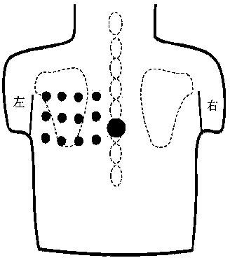
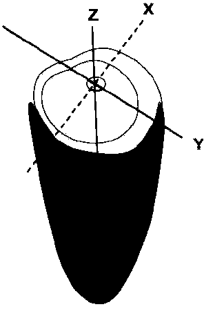
[0041] Figure 1 - Figure 6 The body surface array electrode three-dimensional lead system is demonstrated, which combines the classic three-dimensional ECG vector system and the modern general body surface array electrode that maps the corresponding heart parts (see references 2, 3). The body surface potential obtained through this lead system reflects the transmural potential activity related to the 0-point galvanic couple in the corresponding cardiac anatomical parts (such as the left ventricular middle septum, anterior wall, lateral wall, posterior wall, etc.). Such as Figure 3 to Figure 6 As shown, according to the routine el...


PUM


Abstract

The invention relates to medical examination and diagnosis methods and instruments, in particular to a multi-dimensional electrocardiosignal imaging system and method. The body surface electric potential mapping technology and the electrocardio vector technology are integrated, a new non-invasive platform is provided for recording the heart-across-myocardium electrical activity information, and in other words, a new non-invasive body surface heart-across-myocardium electrical activity information imaging method and technology which are formed by combining the vector center 0 point galvanic coupling and body surface electric potential mapping technology and are similar to satellite positioning are provided. The non-invasive electrocardio function imaging method and technology for detecting heart-across-myocardium electrocardio activity information with the dissection positioning as a basis are provided. According to the multi-dimensional electrocardiosignal imaging system and method, the arrhythmia reason can be determined, the cardiac muscle diseases such as myocardial hypertrophy, cardiomyopathy, myocardial infarction and myocarditis can be diagnosed, and a brand new diagnosis tool is provided for fast diagnosing the heat diseases in real time.

Description

Technical field [0001] The present invention involves a medical examination diagnosis method and instrument, which involves a multi -dimensional e -signal imaging system and method. Background technique [0002] Heart electrical activity is one of the most basic physiological features of the heart.This activity reflects the self -discipline, excitement and electrical conductivity of the heart excitement organization.Many cardiac diseases can cause excitement and conduction abnormality of the heart electricity. Electric activity to detect the heart is an important part of diagnosis and treatment of cardiac diseases.Currently used to detect ECG activity technology can be summarized into the following aspects: [0003] 1.1 One -dimensional heart electrocomputer (ECG): [0004] The electrical activity of the heart is indirectly recorded through the body surface.This activity reflects the de -polarization and compound activity of myocardium in the cardiac cycle.ECG represents the sum ...

Claims


Application Information

Patent Timeline
IPC IPC(8): A61B5/0402
Inventor 雷鸣郝国梁
Owner 河南省贝威科技有限公司
Learn More