
An Evaluation Method of Spectral CT Image Quality

A CT image and evaluation method technology, applied in the field of image processing, to achieve high efficiency

Active Publication Date: 2017-05-03
SOUTHERN MEDICAL UNIVERSITY
View PDF4 Cites 0 Cited by
  • Summary
  • Abstract
  • Description
  • Claims
  • Application Information

AI Technical Summary

Problems solved by technology

[0016] The present invention proposes an efficient evaluation method for energy spectrum CT image quality, which is simple to implement, can quickly determine the best energy point in energy spectrum CT single-energy images, and can well reflect the visual perception results of image quality, so as to To solve the existence of the existing technology is simply to consider the shortcomings of the background noise

Method used


Image

  • An Evaluation Method of Spectral CT Image Quality
  • An Evaluation Method of Spectral CT Image Quality
  • An Evaluation Method of Spectral CT Image Quality

Examples


Embodiment Construction

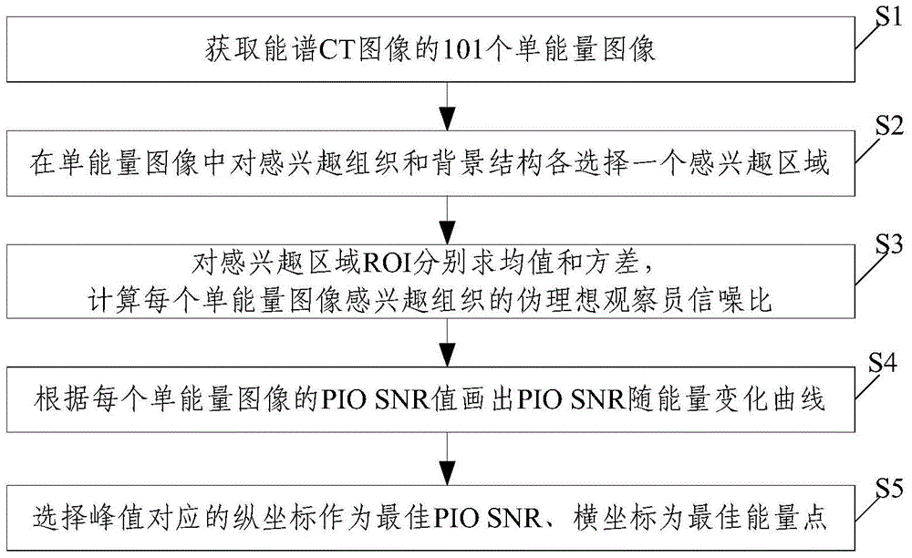
[0038]Specifically, the present invention refers to the optimal value of the ideal observer (Ideal Observer, referred to as IO) to indicate that the scanning protocol of the imaging system is optimal and the amount of data collected by the hardware is maximized, by calculating the pseudo-ideal observer signal-to-noise ratio (Pseudo Ideal Observer Signal-Noise Ratio) of the sampling data. to-Noise ratio (PIO SNR for short) to objectively evaluate spectral CT single-energy images, and finally can quickly find the best energy point showing the tissue of interest from many single-energy images. This method mainly quantitatively analyzes the relationship between the physical factors of imaging and the spatial resolution response of the imaging system, and calculates the PIO SNR value of the region of interest as the basis for evaluating the best energy point. Evaluation, it can evaluate regions of interest such as tissues, organs, and lesions very well, has a strong correlation with...


PUM


Abstract

The invention discloses an energy spectrum CT image quality evaluation method. The method includes the steps of obtaining 101 mono-energy images in an energy range of 40 to 140keV (kiloelectron volt) through energy spectrum CT images; determining an interesting organization structure and a background structure in every mono-energy image and respectively selecting interesting regions ROI of the same area in the interesting organization structure and the background structure; respectively calculating the mean value <t>1 and the variance sigma (2)1 of the interesting region ROI of the interesting organization structure, the mean value <t>0 and the variance sigma (2) 0 of the interesting region ROI of the background structure, and the pseudo ideal observer signal to noise ratio (PIO SNR) of the interesting organization structure of every mono-energy image; drawing a changing curve of the PIO SNR changing with the energy according to the pseudo ideal observer signal to noise ratio (PIO SNR) of every mono-energy image and determining a optimum energy point. The energy spectrum CT image quality evaluation method can quickly determine the optimum energy point of the interesting region, takes the relevance between the background noise and the noise of the interesting region into full consideration, is close to and in line with characteristics of a human visual system, and has the advantages of stability and efficiency.

Description

technical field [0001] The invention belongs to the technical field of image processing, and in particular relates to a method for evaluating the quality of an energy spectrum CT single-energy image. Background technique [0002] In conventional X-ray computed tomography (CT), people still rely on the difference in CT value of substances for judgment, but it is usually difficult to find and identify pathological changes with small differences in CT value in the image , not to mention that the CT value will be affected by many problems such as hardening effect and metal artifact. In order to make up for these deficiencies, physicists have proposed a corresponding solution, that is, energy spectrum CT imaging, by using the energy spectrum characteristics of X-rays and the rich attenuation information of matter. [0003] Physicists use two kinds of X-rays with different energies to scan the object at the same time, and can obtain images of the object under different energy ray...

Claims


Application Information

Patent Timeline
Patent Type & Authority Patents(China)
IPC IPC(8): G06T7/00G06T5/20
Inventor 马建华曾栋高杨黄静
Owner SOUTHERN MEDICAL UNIVERSITY
Learn More