Cutting method of pectoral muscle region in mammary gland X-ray image

A light image and mammary gland technology, applied in the field of medical image processing, can solve problems such as difficult segmentation and inability to accurately locate the pectoralis region

Inactive Publication Date: 2014-04-02
SHENZHEN LANDWIND IND
View PDF6 Cites 20 Cited by
  • Summary
  • Abstract
  • Description
  • Claims
  • Application Information

AI Technical Summary

Problems solved by technology

[0006] The present invention proposes a method for segmenting the pectoralis region in mammography images to solve the technical problem th

Method used


Image

  • Cutting method of pectoral muscle region in mammary gland X-ray image
  • Cutting method of pectoral muscle region in mammary gland X-ray image
  • Cutting method of pectoral muscle region in mammary gland X-ray image

Examples


Embodiment Construction

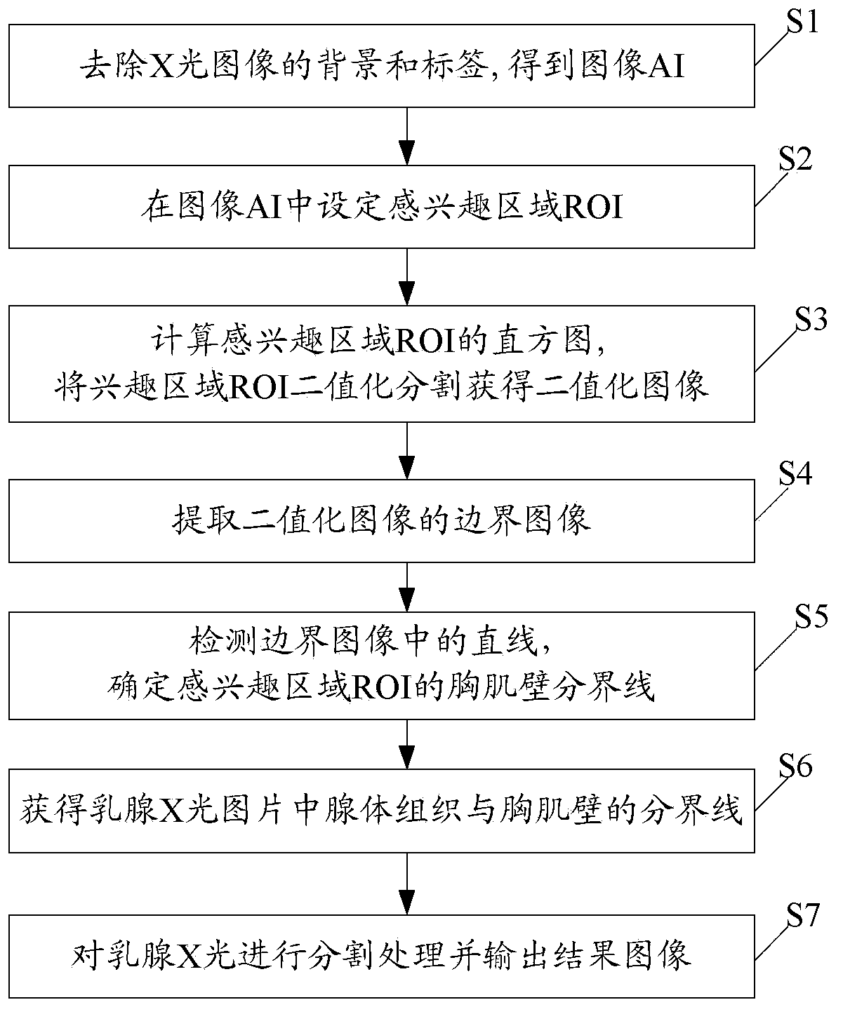
[0025] The present invention removes the background and label of the X-ray image by reading in the X-ray image of the mammary gland using an adaptive threshold value; then, the region of interest (ROI, Region of Interest) is set in the X-ray image of the mammary gland, and the histogram of the ROI is used to calculate Generate a threshold, and generate a black-and-white binary image based on this threshold; then, use the Canny edge operator to extract the boundary image on the black-and-white binary image; finally, find the straight line in the boundary image through Hough transform to determine the mammogram the pectoralis region.

[0026] combine figure 1 As shown, the present invention specifically includes the following implementation steps:

[0027] Step S1, to the input mammogram image (such as Figure 2A shown) for preprocessing to remove the background and labels of the mammogram image, resulting in an image AI containing only tissue regions.

[0028] Mammogram imag...


PUM


Abstract

The invention discloses a cutting method of a pectoral muscle region in a mammary gland X-ray image. The method comprises the steps of fixing an region of interest ROI in a mammary gland X-ray image, performing binarization cutting ROIBW to the ROI, so as to obtain a binarization image ROIBW, extracting an edge image ROIedge of a binarization image ROIBW, detecting a straight line in the edge image ROIedge, fixing a pectoral muscle wall boundary in the ROI, extending the pectoral muscle wall boundary to the boundary of the edge image ROIedge to be intersected with the boundary, obtaining a boundary of glandular tissue and the pectoral muscle wall in the mammary gland X-ray image, and performing cutting processing to the mammary gland X-ray image according to the boundary of the glandular tissue and the pectoral muscle wall. The method fixes the boundary of the glandular tissue and the pectoral muscle wall through the difference of gray features of the glandular tissue and the pectoral muscle wall in the mammary gland X-ray image, and can accurately position the pectoral muscle region to perform image cutting.

Description

technical field [0001] The invention relates to a medical image processing technology, in particular to a method for segmenting a pectoral muscle region in a mammogram. Background technique [0002] The clarity and contrast of the original mammography images are often insufficient. In order to help clinicians better diagnose the disease, it is necessary to process and enhance the original images. In the lateral oblique (MLO, Mediolateral Oblique) image, the breast tissue often contains the pectoralis, which is quite different in nature from the breast area and does not contain calcifications. Information detection and processing according to the unique image features of each region is the premise and important basis for the research of microcalcification and mass detection. The segmentation of pectoralis region plays an important role in the comparison of left and right breasts in mammograms and the delimitation of mass detection regions. At present, the similar related pa...

Claims


Application Information

Patent Timeline
IPC IPC(8): G06T7/00
Inventor 赵明康雨
Owner SHENZHEN LANDWIND IND
Why Eureka
Try Eureka