Orthopedic guide reset pincers

A reduction forceps, orthopedic technology, applied in medical science, surgical forceps, surgery, etc., can solve the problems of failed surgery, insufficient surgical space, inconvenient operation of the guide, reduce the probability of wound infection, and facilitate the surgical incision. Healing, avoidance of iatrogenic injury

Active Publication Date: 2015-05-27
段强民
View PDF8 Cites 0 Cited by
  • Summary
  • Abstract
  • Description
  • Claims
  • Application Information

AI Technical Summary

Problems solved by technology

[0002] When treating humeral intercondylar fractures, humeral supracondylar fractures, humeral medial epicondyle fractures, and lateral epicondyle fractures, the ulnar nerve walks on the posterior side of the medial epicondyle and the radial nerve is in close contact with the crest of the lateral epicondyle of the humerus Due to the reason of the bone surface, the traditional method of performing medial and lateral needle fixation is to perform a combined medial and lateral approach, reduce the fracture under direct vision, and perform blind needle puncture before performing fluoroscopy to confirm the entry and exit points of the Steiner pin and the position on the opposite side. Whether the length is ideal, because a point cannot determine a straight line when threading the needle, the needle cannot be accurately found after the needle is inserted from the entry point. Pain and recovery time, and there is a high risk of iatrogenic damage to important blood vessels and nerves during blind puncture
[0003] Existing orthopedic repositioning forceps only have the function of resetting, which can reset and fix the bone at the fracture site, and cannot guide needles through. The current orthopedic guides, such as the Chinese utility model 200720018312.0 adjustable orthopedic drilling guide, all adopt a rectangular structure. One long side of the rectangle determines the needle entry point and the needle exit point, and the other long side guides. First of all, this structure does not have a reset function. If orthopedic reduction forceps and this guide are used at the same time, there is not enough surgical space, and the surgical wound Secondly, this kind of guide is inconvenient to operate, and the doctor needs to operate it with both hands. When positioning on a smooth bone surface, it is difficult to fix the guide accurately due to the instability of the rectangle; the front side of the rectangular guide guides, The back side is fixed, and the hands are clamped. In actual use, it is found that it is difficult for the doctor to grasp the strength. The guide is easy to loose when the strength is weak, and the fracture is easily dislocated if the strength is too high, which increases the difficulty of the operation and even causes the operation to fail.

Method used


Image

  • Orthopedic guide reset pincers
  • Orthopedic guide reset pincers

Examples


Embodiment Construction

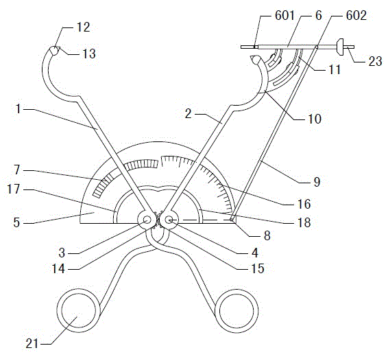
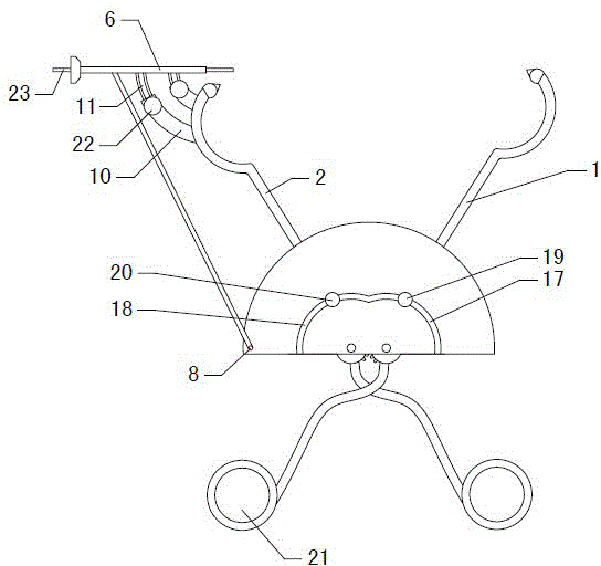
[0030] Figure 1~2 It is the best embodiment of the orthopedic guide reduction forceps of the present invention, below in conjunction with the attached Figure 1~2 The present invention will be further described.

[0031] Refer to attached figure 1 : Orthopedic guide reduction forceps, including left forceps body 1, right forceps body 2, fixing plate 5 and sleeve 6, left forceps body 1 and right forceps body 2 swinging at equal angles are fixed on the plate 5, right forceps body 2 front end A sleeve 6 is provided for swinging, and an adjustment rod 9 is hinged between the right end of the sleeve 6 and the right side of the fixing plate 5. Through the adjustment rod 9, the sleeve 6 always points to the front end of the left clamp body 1, and the fracture fixation is determined through the sleeve 6. direction of the needle.

[0032] The fixed plate 5 is vertically fixed with a left rotating shaft 3 and a right rotating shaft 4, the left pliers body 1 rotates and is fixed on t...


PUM


Abstract

An orthopedic guide reduction forceps belongs to the field of orthopedic medical instruments. The left clamp body (1) is fixed on a fixed plate (5) through the rotation of the left rotating shaft (3), the right clamp body (2) is fixed on the fixed plate (5) through the rotation of the right rotating shaft (4), and the left clamp body (1 ) and the right clamp body (2) are provided with an equal-angle swing mechanism, and a sleeve (6) is provided at the front end of the right clamp body (2). The sleeve (6) rotates around the swing center point (601), and the sleeve ( 6) The right end is provided with a hinge point (602) and an adjustment rod (9) is hinged, and the lower end of the adjustment rod (9) is hinged at the positioning hole (8) on the right side of the fixed plate (5) at the swing center point (601) to The length of the hinge point (602) is equal to the center distance between the positioning hole (8) and the right shaft (4), and the adjusting rod (9) makes the sleeve (6) always point to the front end of the left clamp body (1). The invention has the advantages of accurate guiding and positioning, convenient use, minimally invasive surgical treatment and the like.

Description

technical field [0001] The orthopedic-guided reduction forceps belongs to the field of orthopedic medical devices, and in particular relates to an orthopedic-guided reduction forceps which are used for reducing fractures and guiding fixation pins when treating fractures. Background technique [0002] When treating humeral intercondylar fractures, humeral supracondylar fractures, humeral medial epicondyle fractures, and lateral epicondyle fractures, the ulnar nerve walks on the posterior side of the medial epicondyle and the radial nerve is in close contact with the crest of the lateral epicondyle of the humerus Due to the reason of the bone surface, the traditional method of performing medial and lateral needle fixation is to perform a combined medial and lateral approach, reduce the fracture under direct vision, and perform blind needle puncture before performing fluoroscopy to confirm the entry and exit points of the Steiner pin and the position on the opposite side. Wheth...

Claims


Application Information

Patent Timeline
Patent Type & Authority Patents(China)
IPC IPC(8): A61B17/90A61B17/56
CPCA61B17/29A61B17/88A61B2017/564A61B17/90
Inventor 段强民
Owner 段强民
Why Eureka
Try Eureka