Pure entire foetal genome DNA isolation method

A whole-genome, fetal technology, applied in the medical field, can solve the problems of unidentified fetuses, amniotic membrane rupture, intrauterine death, etc., and achieve the effects of accurate and reliable diagnosis results, short life cycle, and large storage capacity

Inactive Publication Date: 2013-11-06
SHANGHAI GREENGLOBE BIOTECH
View PDF1 Cites 5 Cited by
  • Summary
  • Abstract
  • Description
  • Claims
  • Application Information

AI Technical Summary

Problems solved by technology

[0004] At present, invasive methods are still used in prenatal genetic diagnosis, and amniocentesis and chorionic villi sampling are the two most commonly used methods. However, there are the following risks when the fetus is invasively obtained: 1. Transient fetal bradycardia; 2. 0.1% to 0.9% of the subjects had premature birth or fetal death; 3. Umbilical cord placental bleeding after umbilical cord blood collection; 4. Intra-amniotic infection; 5. Amniotic membrane rupture; 6. Fetal malformation
[0005] Among non-invasive methods, ultrasonography can only distinguish some fetuses with obvious congenital abnormalities morphologically, but cannot distinguish morphologically normal fetuses with other genetic defects
The determination of maternal peripheral serum markers is an indirect detection from maternal serum markers, so the inspection items are limited, and the current screening is mainly for Down's syndrome, which is only one of the chromosomal trisomy syndromes. Most of the genetic diseases are not detectable by similar non-invasive methods

Method used


Image

  • Pure entire foetal genome DNA isolation method

Examples


Embodiment 1

[0030] Example 1, Separation and Obtaining Pure Fetal Complete Genome

[0031] 1.1. Separation of nucleated red blood cells from maternal peripheral blood

[0032] Take 10ml of peripheral venous blood from mothers (pregnant women) from 9 to 38 weeks of pregnancy, anticoagulate with 4mmol / L EDTA, and then dilute 1:1 with phosphate buffered saline (PBS, pH7.4). In a 15ml Falcon centrifuge tube, add 5ml cell separation solution (Sigma Company), slowly add 10ml of the above-mentioned diluted pregnant woman's blood along the wall of the centrifuge tube, so that the blood is covered in the cell separation solution Centrifuge at 400×g for 30 minutes at room temperature. absorbed in plasma and The mononuclear cell layer in between was washed once with PBS buffer (containing 5mmol / L EDTA and 0.5% BSA by volume fraction), approximately every 10 7 A mononuclear cell suspension was prepared by adding 80 μL of PBS buffer (pH 7.4) to each cell.

[0033] Add CD71 immunomagnetic beads...

Embodiment 2-6

[0047] Embodiment 2-6, application

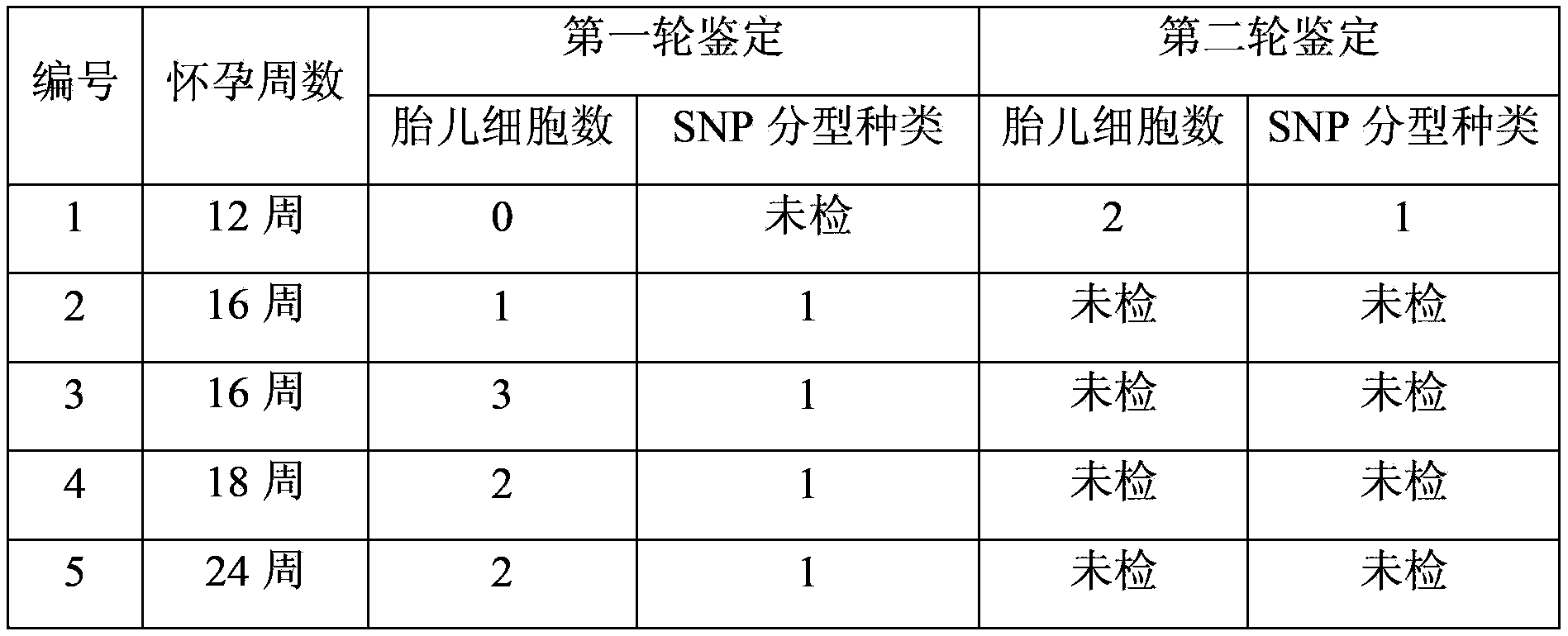
[0048] According to the method described in Example 1, 5 pregnant women who were pregnant for 12-24 weeks were selected, and 20 ml of peripheral blood from the pregnant women were respectively collected to separate nucleated red blood cells. 10 nucleated erythrocytes isolated from each pregnant woman were selected for whole-genome amplification, and then individual identification was carried out with the mother's somatic cells as a control to determine the fetal genome in the amplified genome. Among them, 4 pregnant women identified 1 -3 fetal cells, 1 pregnant woman was not detected for the first time. Another 10 nucleated erythrocytes were taken from the undetected pregnant woman for the second round of whole-genome amplification and identification, and 2 of them were found to be fetal cells. The above 5 volunteers only identified the SNP type of one fetus, which means that there is only one fetus. Since then, the fetuses have been born ...


PUM


Abstract

Provided is a method for isolating and obtaining pure and complete fetus genome DNA, comprising the following steps: isolating a nucleated erythrocyte from the maternal (pregnant woman) peripheral blood, identifying the individual hereditary feature subtype of the mother and / or father of the fetus; conducting high fidelity whole genome DNA amplification on the signal cell isolated from the nucleated erythrocyte; identifying the individual hereditary feature subtype of the fetus according to the laws of hereditary, and determining a fetus-originating genome DNA. The fetus-originating pure whole genome DNA provided by the present invention lays a material foundation for prenatal heredity diagnosis.

Description

technical field [0001] The invention belongs to the field of medicine, and in particular relates to a method for separating and obtaining pure fetal complete genome DNA. Background technique [0002] Prenatal genetic diagnosis is based on genetic counseling, mainly through genetic testing and imaging examinations, to make a clear diagnosis of high-risk fetuses, so that people can make appropriate choices or treatments during pregnancy on a scientific basis, thereby reducing the risk of birth. Defect rate, improve eugenic quality and population quality. [0003] Prenatal genetic diagnosis includes both invasive and noninvasive types. The former mainly includes amniocentesis, chorionic villus sampling, umbilical cord blood sampling, fetoscopy and embryo biopsy, etc., while the latter includes ultrasonography, measurement of maternal peripheral serum markers, non-invasive fetal gene and cytogenetic analysis, etc. [0004] At present, invasive methods are still used in prenata...

Claims


Application Information

Patent Timeline
IPC IPC(8): C12N15/10
CPCC12Q1/68C12N15/10C12Q1/6806
Inventor 蔡勇平潘星华
Owner SHANGHAI GREENGLOBE BIOTECH
Why Eureka
Try Eureka