
Breast ultrasonoscopy automatic segmentation method based on mean shift and divide

A mean-shift, ultrasound image technology, applied in the field of image processing, can solve the problems of low algorithm robustness, time-consuming processing, sensitivity to speckle noise, etc., and achieve the effect of avoiding manual interaction, high level of automation, and easy implementation.

Inactive Publication Date: 2013-09-11
BEIJING UNIV OF TECH +1
View PDF1 Cites 13 Cited by
  • Summary
  • Abstract
  • Description
  • Claims
  • Application Information

AI Technical Summary

Problems solved by technology

These methods have some major disadvantages: (1) most methods require manual interaction, such as manual selection of seed points or initial contours; (2) most methods are sensitive to speckle noise, and because ultrasound images have low contrast and tissue-related textures, so Accurate segmentation is difficult; (3) Most methods have high algorithm complexity and long processing time, which are difficult to meet clinical requirements
However, these methods have the following problems: (1) The algorithms for automatically finding seed points or regions of interest are often not robust, and are only effective for some images, which affects the accuracy of automatic segmentation; (2) The algorithm complexity is high, and processing Time-consuming, it is difficult to meet the clinical speed requirements for automatic segmentation
[0005] The watershed algorithm is a fast segmentation method widely used in image segmentation, but due to the inherent characteristics of ultrasound images, too many regions will be generated during the submersion process, which will lead to slow segmentation and low accuracy.

Method used


Image

  • Breast ultrasonoscopy automatic segmentation method based on mean shift and divide
  • Breast ultrasonoscopy automatic segmentation method based on mean shift and divide
  • Breast ultrasonoscopy automatic segmentation method based on mean shift and divide

Examples


Embodiment Construction

[0038] The extraction process is described in detail with reference to the accompanying drawings and practical examples. The data used are 25 ultrasound images of clinical breast tumors collected by Xinbo medical mammography ultrasound imaging system. The following is a step-by-step introduction:

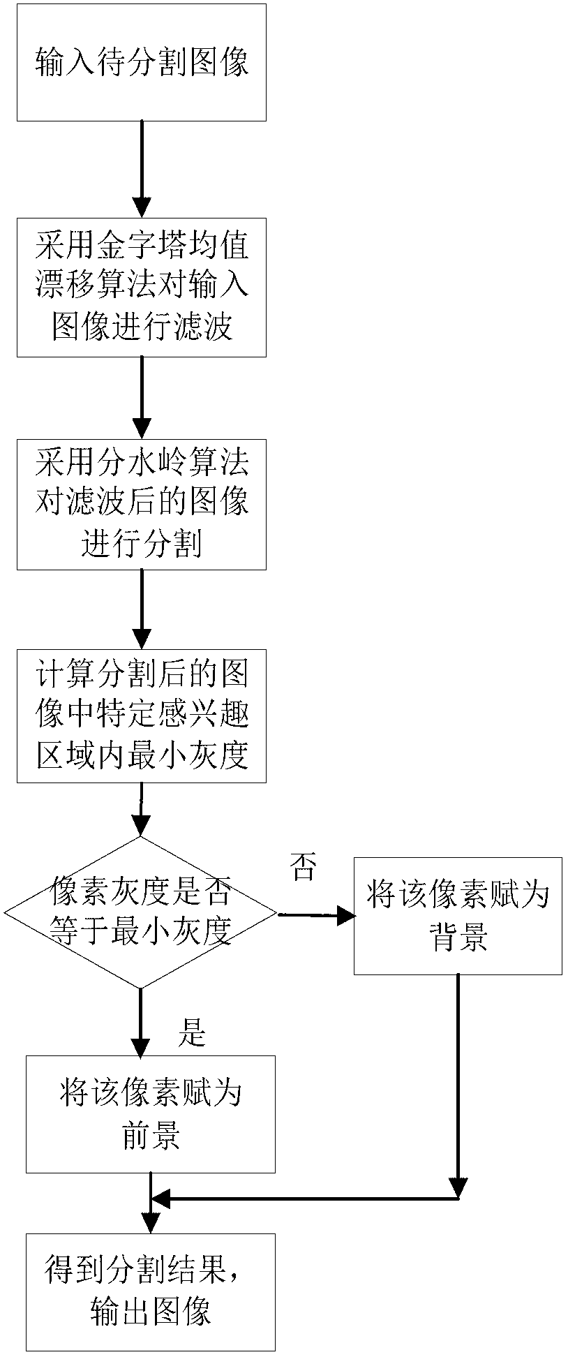
[0039] 1. Use the pyramid mean shift algorithm to process an original breast tumor ultrasound image I to be segmented (such as Figure 4 Shown) to filter, get the filtered image I f . The basic steps of the pyramid mean shift algorithm are as follows: figure 2 shown. The effect picture after filtering is as follows Figure 5 shown. The specific implementation steps are as follows:

[0040] (1). Gaussian pyramid decomposition of the highest layer number L is performed on the breast tumor ultrasound image I, L≥2, and the L-layer image I is obtained 1 ,...,I L , image I L the base of the pyramid;

[0041] (2). For layer L image I L Perform mean shift filtering to obtain th...


PUM


Abstract

The invention discloses a breast tumor ultrasonoscopy automatic segmentation method based on mean shift and divide. A breast tumor ultrasonoscopy is filtered by means of a pyramid mean shift algorithm, the filtered breast tumor ultrasonoscopy is segmented by means of a divide algorithm, the minimum grey level of a specific area of interest in the breast tumor ultrasonoscopy which is segmented through the divide algorithm is calculated according to the experiential knowledge that tumors are generally located on the middle portion or the upper portion of the ultrasonoscopy and the average image intensity is low, the ultrasonoscopy which is segmented through the divide algorithm is traversed, a pixel is regarded as a foreground if the grey level of the pixel is equal to the minimum grey level, the pixel is regarded as a background if the grey level of the pixel is not equal to the minimum grey level, and thus a target tumor area is obtained, namely a final binary image of the tumor segmented result. By means of the breast tumor ultrasonoscopy automatic segmentation method based on mean shift and divide, the boundary of a tumor in the breast tumor ultrasonoscopy is clear and can be retrieved automatically, and the breast tumor ultrasonoscopy can be segmented fast, accurately and automatically.

Description

technical field [0001] The invention belongs to the technical field of image processing, and in particular relates to a method for automatic segmentation of medical ultrasonic images, which is a reformed technology for automatic segmentation of ultrasonic images of breast tumors by mean shift and watershed algorithm. Background technique [0002] Breast cancer is one of the most common malignant tumors in women. At present, the most effective detection and diagnosis method is mammography. However, the low specificity of mammography has resulted in a large number of unnecessary biopsies, which not only brings pain to patients, but also increases costs. In addition, ionizing radiation from mammography poses health risks to patients and physicians. Ultrasound imaging has the characteristics of low cost, non-invasiveness, and real-time performance, and has become one of the important methods for breast tumor detection. However, breast ultrasound images have low contrast, speck...

Claims


Application Information

Patent Timeline
IPC IPC(8): G06T7/00
Inventor 吴水才周著黄林岚赵磊张晓春王宇龙
Owner BEIJING UNIV OF TECH
Learn More