
Method of inspecting target stone with B ultrasound of extracorporeal shock wave lithotripter

An extracorporeal shock wave and lithotripsy technology, applied in echo tomography, organ movement/change detection, ultrasound/sonic/infrasonic Permian technology, etc., can solve problems such as low efficiency, improve treatment effect, and facilitate promotion , reduce the effect of surgical injury

Active Publication Date: 2013-06-19
SHENZHEN HYDE MEDICAL EQUIP
View PDF2 Cites 2 Cited by
  • Summary
  • Abstract
  • Description
  • Claims
  • Application Information

AI Technical Summary

Problems solved by technology

[0003] Aiming at the technical problems existing in the prior art, the technical problem to be solved by the present invention is to overcome the inefficiency of the shock wave triggered by the timing trigger or the heart rate synchronously triggering on the calculi, and to provide a method for machine identification of calculi by using B-ultrasound images. The method of breathing back to the focus of the wave source ensures that the impact force from the wave source can accurately act on the stones

Method used


Image

  • Method of inspecting target stone with B ultrasound of extracorporeal shock wave lithotripter
  • Method of inspecting target stone with B ultrasound of extracorporeal shock wave lithotripter
  • Method of inspecting target stone with B ultrasound of extracorporeal shock wave lithotripter

Examples


Embodiment Construction

[0019] The invention will be described in detail below in conjunction with the accompanying drawings.

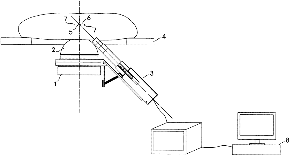
[0020] like figure 1 As shown, the equipment extracorporeal shock wave lithotripter adopting the inventive method comprises a shock wave source 1, a water bag 2, a B-ultrasound probe 3, an operating bed 4, and the B-ultrasound probe 3 is connected to a B-ultrasound machine 9, and the B-ultrasound machine 9 is connected to a control computer 8 . The cut plane of the B-ultrasound probe 3 intersects the central axis of the wave source in the shock wave source 1 at the focus 5 of the wave source. When the stone drifts out of the wave source focus 5, there is no or very little stone image in the section image of the B-ultrasound probe 3, such as the stone 7 drifting out of the wave source focus. When the stone returns to the wave source focus 5, the B-ultrasound probe 3 A stone image will appear in the section image, such as a stone located at the focal point of the wave source...


PUM


Abstract

The invention relates to a method of inspecting target stone with B ultrasound of an extracorporeal shock wave lithotripter. The method comprises the following steps: a. moving a section of a B ultrasound probe to intersect with the axle wire of a shock wave cup at a focus of a wave source, namely, the point is the focus of the wave source of shock waves; b. transmitting the scanned images collected by the B ultrasound probe in real time to a computer, and setting a scanned image with stone located in the focus of the wave source as a template image; c. comparing the images scanned by the B ultrasound probe in real time with the template image through the computer, starting the shock waves to break the stone if the images scanned by the B ultrasound probe in real time are matched with the template image, then skipping to a step d, otherwise, not starting the shock waves to break the stone, and then skipping to the step c; d. automatically collecting a B ultrasound section image which is shocked and with the stone still located in the focus of the wave source to replace the former template image through the computer, and then skipping to the step c. The method of inspecting the target stone with the B ultrasound of the extracorporeal shock waves lithotripter has the advantages that by setting a threshold value to judge whether the stone shifts to the focus of the wave source or not, the shock waves emitted by a machine are guaranteed to act on the stone, a treatment effect is improved, operation damage of a patient is reduced, and the method of inspecting the target stone with the B ultrasound of the extracorporeal shock wave lithotripter is convenient to popularize.

Description

technical field [0001] The invention relates to the field of extracorporeal shock wave lithotripter medical equipment, in particular to a method for B-ultrasonic detection of target stones by an extracorporeal shock wave lithotripter. Background technique [0002] Extracorporeal shock wave lithotripter is widely used in our country. Its positioning principle mainly relies on B-ultrasound or X-ray images to find stones, and moves human stones to the wave source focus of the wave source through the relative movement of the wave source and the bed, and finally uses the wave source to converge the impact force on the stones to crush the stones. However, in the actual lithotripsy process, B-ultrasound images show that the stones drift back and forth about 2cm with the patient’s breathing relative to the focus of the wave source, so that only about 30% of the shock waves triggered by timing can act on the stones, and the rest of the shock waves will act on the stones. In other pl...

Claims


Application Information

Patent Timeline
Patent Type & Authority Applications(China)
IPC IPC(8): A61B8/08A61B8/14A61B17/225
Inventor 朱恒宇朱伟辉陈文韬高田桂孙西钊
Owner SHENZHEN HYDE MEDICAL EQUIP
Learn More