Noninvasive method for determining iron content in brain tissue

A technology of iron content and brain tissue, which is applied in the directions of diagnostic recording/measurement, medical science, sensors, etc., can solve the problems of inaccurate measurement of brain regions and the influence of different parameters, and achieve the effect of simple operation process.

Inactive Publication Date: 2013-11-27
ZHEJIANG UNIV
View PDF4 Cites 1 Cited by
  • Summary
  • Abstract
  • Description
  • Claims
  • Application Information

AI Technical Summary

Problems solved by technology

[0003] In order to overcome the limitations of some specific brain regions that exist in the existing measurement of brain iron content indicators, such as inaccurate measurement, being affected by different parameters, and requiring specific workstations, the purpose of the present invention is to provide a method for non-invasive measurement of brain tissue iron content

Method used


Image

  • Noninvasive method for determining iron content in brain tissue
  • Noninvasive method for determining iron content in brain tissue

Examples


Embodiment

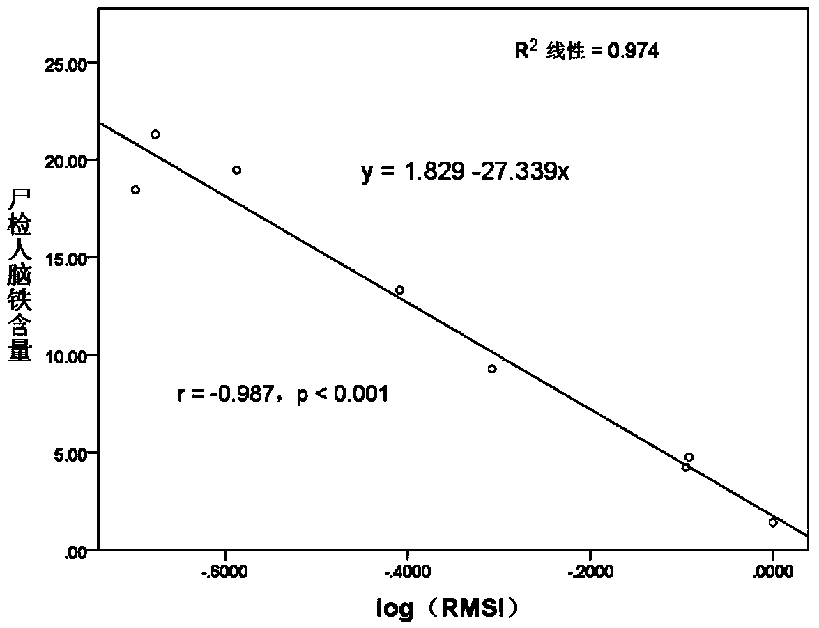
[0030] 1) 30 healthy people aged 30-100 years were recruited and signed the informed consent form.

[0031] 2) Using a magnetic resonance apparatus (Signa Excite HD, General Electric Medical System, Milwaukee, USA) produced by GE with a magnetic field strength of 3.0 Tesla, the participants were subjected to multi-echo acquisition of T2*-weighted three-dimensional gradient echoes of the whole brain. wave sequence (Enhanced gradient echo T2* weighted angiography, ESWAN) and save the magnitude images in the resulting image data. The parameters set are as follows: 3D multi-echo gradient echoes are used, a total of 11 equally spaced echoes: first echo time = 4.5 ms, echo interval time = 4.5 ms, repetition time = 58 ms, deflection Angle = 20°, Matrix = 256 × 256, Field of View = 240 × 240 mm2, Layer Thickness = 2.0 mm, Layer Spacing 0 mm, Resolution = 0.4688 × 0.4688 mm / pixel.

[0032] 3) Measure the signal intensity of caudate nucleus 1, putamen 2, globus pallidus 3, thalamus 4, ...


PUM


Abstract

The invention discloses a noninvasive method for determining iron content in brain tissue. The method comprises the steps of performing whole brain magnetic susceptibility imaging on participator with magnetic resonance instrument, and storing amplitude image in obtained image data; measuring average signal intensities of caudate nucleus, putamen, globus pallidus, thalamus, red nucleus, substantia nigra, frontal lobe white matter and medulla oblongata in brain region of each participator, and dividing with the average signal intensity of medulla oblongata as internal reference respectively, to obtain relative amplitude signal intensity data; performing normal distribution conversion on the obtained relative amplitude signal intensity data, performing Pearson linear correlation analysis with brain region iron content distribution data of population of same age group published by autopsy research, and fitting to obtain a simple relation equation according to correlation analysis result. The method can non-invasively accurately estimate iron content in brain, only needs measurement and calculation on a simple third-party image processing software during measurement, and needs no special workstation.

Description

Technical field [0001] The present invention involves a method of non -invasive measurement of brain iron content in medical nerve images. Background technique [0002] Iron deposition in the brain is related to certain neurodegenerative diseases. With the development of medical imaging, many magnetic resonance parameters are used to evaluate the iron tissue iron content.The relaxation rate (R2*), quantitative magnetic sensitivity, etc.However, there are certain defects for these parameters to reflect the iron content: if the phase value is directional due to the existence of the phase information, the iron content in some specific brain areas (such as pale balls) is not accurate; R2*also includes the same time includes the same containing includes containing it.The information of the magnetic field and brain tissue, different tissue components have a certain impact on their values; at the same time, various magnetic resonance parameters, such as Echo Time (TE), repetition time, ...

Claims


Application Information

Patent Timeline
Patent Type & Authority Patents(China)
IPC IPC(8): A61B5/055
Inventor 楼敏严慎强孙建忠
Owner ZHEJIANG UNIV
Why Eureka
Try Eureka