Medical image fusion method based on multidirectional empirical mode decomposition

An empirical mode decomposition and medical image technology, applied in image enhancement, image data processing, instruments, etc., can solve problems such as image local position distortion, doctor misdiagnosis, and lack of detail information, so as to reduce impact, improve quality, and strengthen detail information The effect of gaining power

Inactive Publication Date: 2014-04-16
HENAN UNIV OF SCI & TECH
View PDF4 Cites 0 Cited by
  • Summary
  • Abstract
  • Description
  • Claims
  • Application Information

AI Technical Summary

Problems solved by technology

But no matter what kind of wavelet it is based on, there is a problem in image fusion: the fused image will be distorted in a local position, which is acceptable in general applications, but in medical images, this may cause doctors Misdiagnosis affects the patient's follow-up treatment
However, the traditional two-dimensional empirical mode decomposition has defects: there are dark spots in the intrinsic mode component image obtained from the decomposition
[0006] In summary, there are still some deficiencies in the existing fusion technology applied to medical image fusion: the fusion image based on wavelet and ultrawavelet will appear local distortion, and the dark spots of the intrinsic mode component image decomposed by the traditional empirical mode decomposition method are all right. The fusion result has a great influence. In the fusion graph decomposed by the window empirical mode, a small amount of detail information will be missing, and the quality of the fusion rule also has a huge impact on the fusion effect.

Method used


Image

  • Medical image fusion method based on multidirectional empirical mode decomposition
  • Medical image fusion method based on multidirectional empirical mode decomposition
  • Medical image fusion method based on multidirectional empirical mode decomposition

Examples


Embodiment Construction

[0043] In order to better understand the technical solutions of the present invention, the implementation manners of the present invention will be described in detail below in conjunction with the accompanying drawings.

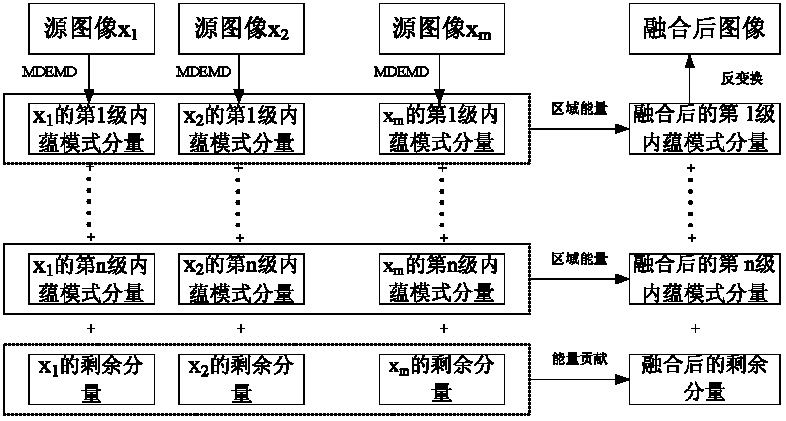
[0044] Such as figure 1 As shown, the present invention first processes the source image x 1 , x 2 ,...x m Carry out multi-directional empirical mode decomposition to obtain n-level intrinsic mode components and a residual component of each source image, and perform fusion processing on all levels of intrinsic mode components according to the regional energy rule; use energy contribution rules for the low-frequency residual components of the source image Carry out fusion processing; finally inverse transform to obtain the fusion image.

[0045] The concrete implementation of the present invention is as follows:

[0046] 1. Use the MDEMD algorithm to match the source image x to be fused 1 , x 2 ,...x m Carry out the decomposition of the same series resp...


PUM


Abstract

The invention relates to a medical image fusion method based on multidirectional empirical mode decomposition. The method comprises the following steps of: performing the multi-scale and multidirectional decomposition on the collected medical source images in different varieties by adopting the multidirectional empirical mode decomposition, acquiring an intrinsic mode component of a multi-scale and multidirectional high-frequency component of a source image; performing the fusion treatment according to the local energy rule in order to effectively extract the high-frequency detailed information of every source image; performing the fusion treatment on the residual low-frequency component of the source image by adopting the energy contribution rules; and finally, inversely transforming to obtain the fused image. The fused image can effectively improve the visual effect of the fused image and avoid the shortcomings of partial distortion or deletion of the fusion image caused by small wave, ultra-small wave and window empirical mode decomposition fusion algorithm; the parameter does not need to be selected manually, the detailed information of the source image can be well extracted, and the self-adaptive image fusion based on a brand-new multi-scale decomposition structure is achieved; the medical image fusion method based on the multidirectional empirical mode decomposition has complete adaptability driven by data and stronger ability of obtaining the detailed information.

Description

technical field [0001] The invention belongs to the technical field of image fusion, and relates to a medical image fusion method based on multidirectional empirical mode decomposition. Background technique [0002] Image fusion is an information fusion technology whose main research content is image. It is to combine two or more images into one image to obtain a more accurate, comprehensive and reliable image description of the same scene. Image fusion technology effectively utilizes the redundancy and complementarity between multiple images to make the fused image more suitable for the human visual system and the needs of computer understanding, analysis and subsequent processing. [0003] Medical image fusion is an important application in image fusion. At present, medical imaging equipment mainly includes: computer tomography (Computer Tomography, CT), magnetic resonance imaging (Magnetic Resonance Imaging, MRI) or the newer positron emission tomography (Positron Emissi...

Claims


Application Information

Patent Timeline
Patent Type & Authority Patents(China)
IPC IPC(8): G06T5/50
Inventor 梁灵飞平子良普杰信黎蔚黄涛
Owner HENAN UNIV OF SCI & TECH
Why Eureka
Try Eureka