
Multimodal molecular imaging monitoring method for ischemia model

A molecular imaging and multi-modal technology, applied in diagnostic recording/measurement, medical science, computerized tomography scanner, etc., can solve problems such as inability to intuitively display the three-dimensional space position information of the light source in the body of small animals, inconvenience of continuous observation, and execution

Active Publication Date: 2016-08-17
FOURTH MILITARY MEDICAL UNIVERSITY +1
View PDF1 Cites 0 Cited by
  • Summary
  • Abstract
  • Description
  • Claims
  • Application Information

AI Technical Summary

Problems solved by technology

However, currently, methods for monitoring the intervention effect of animal models mainly include in vitro immunohistochemistry, immunofluorescence and other means. For the survival of stem cells in small animals, it is necessary to transplant stem cells derived from transgenes with green fluorescent protein (GFP). From animal stem cells to injured animal models, the survival of stem cells needs to be sacrificed and obtained from tissues to observe green fluorescent protein under a microscope for identification (Katstural Jetal, CirculationResearch, 2005). imaging, BLI) technology to observe stem cells expressing luciferase (Cao, F. et al. In vivo visualization of embryonic stem cell survival, proliferation, and migration after cardiac delivery. Circulation113, 1005-1014, 2006); biological Luminescence imaging technology can only obtain two-dimensional plane images, and cannot intuitively display the three-dimensional space position information of the light source in the small animal body, which has great limitations in the visualization effect; in terms of quantitative analysis, bioluminescence imaging can only be used Relative quantitative analysis does not have the meaning of absolute quantification, can refer to: R.Weissleder and M.J.Pittet, "Imaging in the era of molecular oncology," Nature 452,580-589 (2008). In addition, to small animal ischemia model also needs to observe For the angiogenesis after stem cell intervention, the traditional method is to take the tissue of the ischemic area to stain the marker molecule CD31 of the vascular endothelium with an antibody, and compare the angiogenesis between the control group and the intervention group
This method needs to kill the animal model at different stages, which brings great inconvenience to the continuous observation of the experiment

Method used


Image

  • Multimodal molecular imaging monitoring method for ischemia model
  • Multimodal molecular imaging monitoring method for ischemia model
  • Multimodal molecular imaging monitoring method for ischemia model

Examples


Embodiment Construction

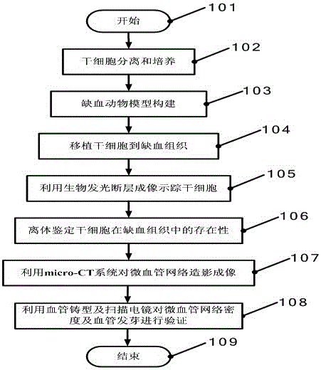
[0028] The present invention proposes a systematic observation method for small animal ischemia model research, and the specific steps are as follows:

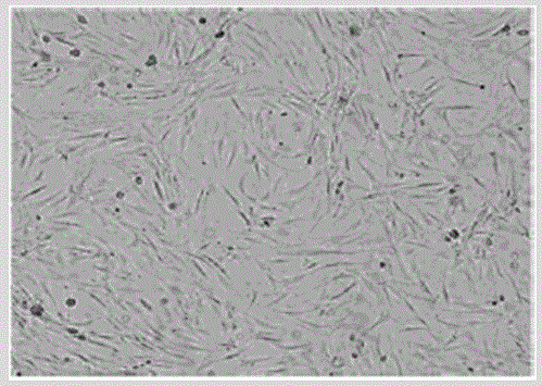
[0029](1) Isolation and culture of stem cells stably expressing luciferase (luciferase) and green fluorescent protein (GFP), stem cells are isolated from transgenic animals stably expressing luciferase (luciferase) and green fluorescent protein (GFP); Because of the stable expression of luciferase, bioluminescence is produced under the action of substrate luciferin, oxygen and adenosine triphosphate (ATP). This bioluminescence can be detected by optical molecular imaging equipment after penetrating biological tissues. Intensity (or the total energy obtained by rebuilding the light source mentioned in the present invention) directly reflects the distribution density or the number of stem cells labeled with luciferase;

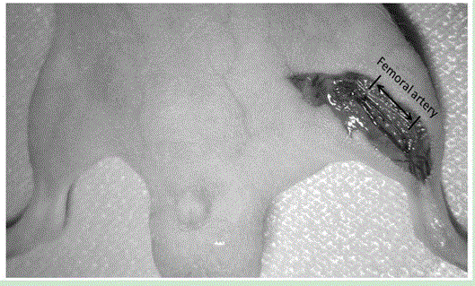
[0030] (2) To construct an ischemic animal model, it is necessary to ensure that the effect of ischemia neith...


PUM


Abstract

The invention relates to the technical field of multimodal molecular imaging, in particular to a multimodal molecular imaging monitoring method for an ischemia model. Through stem cell tracing, angiogenesis observation methods and verification means, it can be confirmed that the bioluminescence tomography method and micro-CT angiography method adopted in the present invention in vivo can be verified through the survival of stem cells, three-dimensional positioning quantitative observation and microvascular network The changes in the density of the blood vessels are used to analyze the situation of angiogenesis. This method avoids the disadvantage of sacrificing small animal models at different time points, and can observe animals continuously for a long time.

Description

1. Technical field: [0001] The invention relates to the technical field of multimodal molecular imaging, in particular to a multimodal molecular imaging monitoring method for an ischemia model. 2. Background technology: [0002] In recent years, small animal molecular imaging technology based on ischemic type has received extensive attention, and studies on stem cell-mediated ischemic injury repair and angiogenesis in this animal model have become commonplace. However, currently, methods for monitoring the intervention effect of animal models mainly include in vitro immunohistochemistry, immunofluorescence and other means. For the survival of stem cells in small animals, it is necessary to transplant stem cells derived from transgenes with green fluorescent protein (GFP). From animal stem cells to injured animal models, the survival of stem cells needs to be sacrificed and obtained from tissues to observe green fluorescent protein under a microscope for identification (Katst...

Claims


Application Information

Patent Timeline
Patent Type & Authority Patents(China)
IPC IPC(8): A61B5/00A61B6/03
CPCA61B6/508
Inventor 曹丰刘俊廷田捷梁继民范伟伟
Owner FOURTH MILITARY MEDICAL UNIVERSITY
Learn More