Positron emission tomography and optical tissue imaging
A technology of positron emission and tomography, which is applied in the field of combined PET and optical imaging systems, can solve the problems of increasing morbidity, prolonging the time of being anesthetized, increasing the time of surgery, etc., so as to reduce the total time and reduce the number of times Effect
- Summary
- Abstract
- Description
- Claims
- Application Information
AI Technical Summary
Problems solved by technology
Method used
Image
Examples
Embodiment Construction
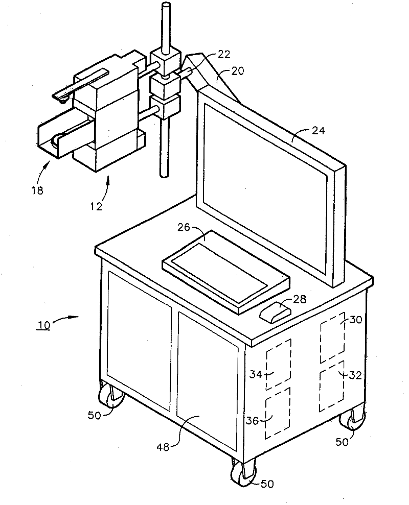
[0010] Positron emission tomography with F-18 fluorodeoxyglucose (FDG) has proven to be a valuable tool for imaging various cancers. Many carcinomas display increased glucose metabolism compared to normal tissues. When FDG is injected intravenously, the FDG concentrates in the tumor so that the tumor can be imaged. In a protocol used in conjunction with the imaging system of the present invention, the patient is injected with a small amount of positron-emitting radiopharmaceutical, such as FDG, one hour before surgery. When the surgeon has removed the cancerous portion, he or she will use dye to mark the resected tissue and the operating table to map the orientation for analysis of the margins.
[0011] The positron emission tomography (PET) tissue sample imager of the present invention uses a pair of small PET cameras to image radiotracer uptake in excised tissue samples. Cancerous tissue is typically identified as one or more focal regions of increased radiotracer accumula...
PUM
 Abstract
Description
Claims
Application Information
 - R&D
- Intellectual Property
- Life Sciences
- Materials
- Tech Scout
- Unparalleled Data Quality
- Higher Quality Content
- 60% Fewer Hallucinations
Browse by: Latest US Patents, China's latest patents, Technical Efficacy Thesaurus, Application Domain, Technology Topic, Popular Technical Reports.
© 2025 PatSnap. All rights reserved.Legal|Privacy policy|Modern Slavery Act Transparency Statement|Sitemap|About US| Contact US: help@patsnap.com


