Method for preparing monocyte type stem cell and application
A mononuclear cell, stem cell technology, applied in the field of cell separation, to reduce the risk and spread of disease
- Summary
- Abstract
- Description
- Claims
- Application Information
AI Technical Summary
Problems solved by technology
Method used
Image
Examples
Embodiment 1
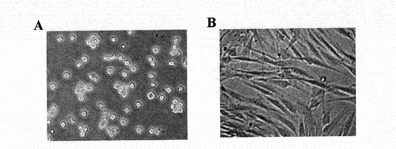
[0040] Example 1 Isolation and cultivation of monocyte-type stem cells from peripheral blood.
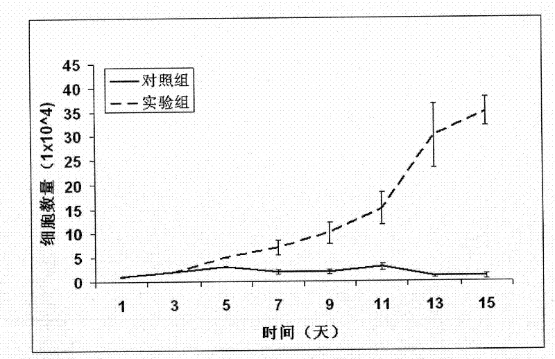
[0041] 200 ml of peripheral blood was extracted, diluted with RPMI 1640 to an equal volume, added with Ficoll-Hypaque, and then centrifuged (550 g) in a Beckmann centrifuge for 30 minutes at 4°C. The harvested buffy coat was washed 2-3 times with RPMI 1640. These cells can be stored immediately in liquid nitrogen or cultured to isolate monocytes. According to the concentration of 2 million cells / milliliter, after adding the culture medium to dilute, transfer the cells to the culture dish for 8-12 hours (37°C, 8% CO2), remove the floating cells, wash 5 times with RPMI1640 medium. Add 5-10 ml of RPMI 1640 medium containing 10% calf serum, and forcefully wash the cells attached to the culture dish to obtain 90-95% pure monocytes. 99% pure monocytes can be further obtained by antibody-binding magnetic bead method. When monocytes were cultured for 5 days by adding 50 ng / ml of M-CSF, 1...
Embodiment 2
[0043] Example 2 Differentiation of macrophages, T lymphocytes and NK cells
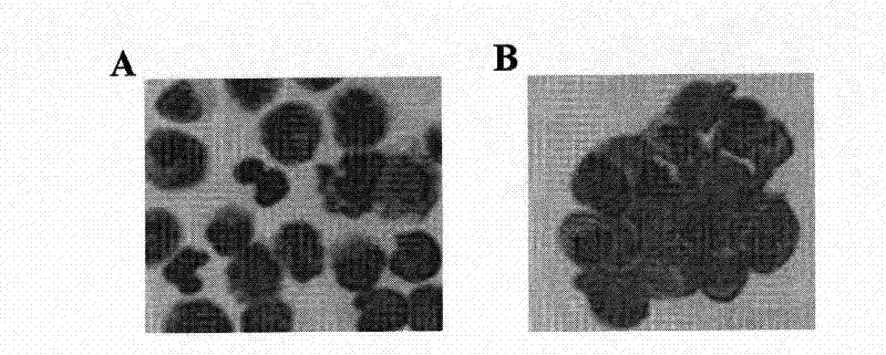
[0044] In order to confirm that monocyte-type stem cells have the ability to differentiate into T lymphocytes, monocytes treated with 50 ng / ml M-CSF, 1000 activity units / ml LIF, and 20 ng / ml IL-6 for 14 days Add 5 ng / ml lipopolysaccharide (macrophage activating factor) for two days. The monocyte-type stem cells thus treated are converted into macrophages. This shift was confirmed by the following experiments, morphological features, increases in HLA-DR, HLA-DQ, immunostaining for IL-10 and TNF-a ( image 3 , Figure 4 ).
[0045] In order to confirm that monocyte-type stem cells have the ability to differentiate into T lymphocytes, monocytes treated with 50 ng / ml M-CSF, 1000 activity units / ml LIF, and 20 ng / ml IL-6 for 14 days Add 1200 activity units / ml of IL-2 (T lymphocyte activating factor) and culture for 4 days. The monocyte-type stem cells thus treated are transformed into T lymphocytes. ...
Embodiment 3
[0047] Example 3 Epithelial cell differentiation
[0048]In order to confirm that monocyte-type stem cells have the ability to differentiate into epithelial cells, monocytes treated with 50 ng / ml M-CSF, 1000 activity units / ml LIF, and 20 ng / ml IL-6 for 14 days were added 100 ng / ml epidermal growth factor (EGF), cultured for 4 days. 75% of the monocyte-type stem cells thus treated showed an epithelial morphology, 71 ± 4% of the cells showed an epithelial morphology after immunostaining, and 68 ± 5% of the cells were positive for E-cadherin, both proteins of which are epithelial marker protein. In the control group, only 4±1% pan-keratins were positive, and 3±2% E-cadherin was positive ( Figure 6 ). Therefore, the monocyte-type stem cells of the present invention can be induced to differentiate into other types of cells other than blood cells. Suitable differentiation-inducing doses can be determined by those of ordinary skill in the art using known routine techniques.
PUM
 Abstract
Description
Claims
Application Information
 - Generate Ideas
- Intellectual Property
- Life Sciences
- Materials
- Tech Scout
- Unparalleled Data Quality
- Higher Quality Content
- 60% Fewer Hallucinations
Browse by: Latest US Patents, China's latest patents, Technical Efficacy Thesaurus, Application Domain, Technology Topic, Popular Technical Reports.
© 2025 PatSnap. All rights reserved.Legal|Privacy policy|Modern Slavery Act Transparency Statement|Sitemap|About US| Contact US: help@patsnap.com


