
In-situ data collection architecture for computer-aided diagnosis

A technology of measured data and medical data, applied in the field of data collection, it can solve problems such as difficulty in obtaining enough data or known cases, high cost, and the reluctance of hospitals to disclose

Active Publication Date: 2007-10-24
KONINKLIJKE PHILIPS ELECTRONICS NV
View PDF0 Cites 7 Cited by
  • Summary
  • Abstract
  • Description
  • Claims
  • Application Information

AI Technical Summary

Problems solved by technology

[0003] One of the main problems with CAD is the difficulty in obtaining enough data or known cases to train a computer
There are many reasons besides technical difficulties, such as hospitals' reluctance to release images of patients, the high cost of obtaining such data, or other social / political reasons

Method used


Image

  • In-situ data collection architecture for computer-aided diagnosis
  • In-situ data collection architecture for computer-aided diagnosis
  • In-situ data collection architecture for computer-aided diagnosis

Examples


Embodiment Construction

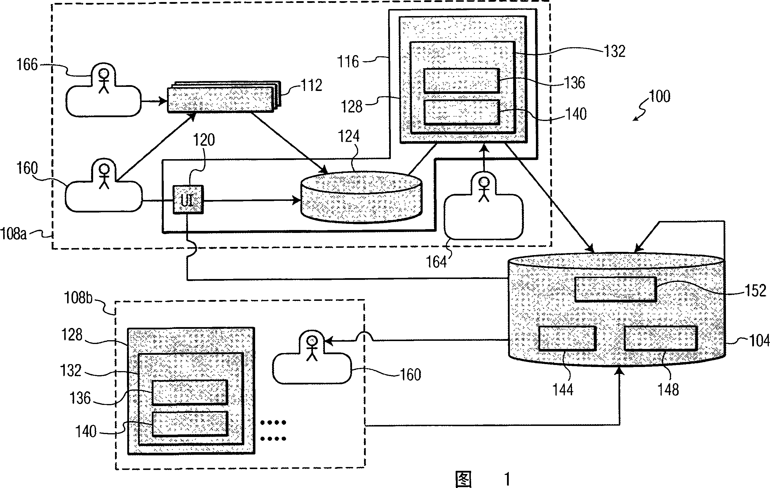
[0017] FIG. 1 depicts, by way of an illustrative and non-limiting example, a CAD input information collection system 100 according to the present invention. System 100 includes diagnostic decision support server 104 and customer site hospitals (or customer sites) 108a and 108b. There may be only one client site hospital or more than two client site hospitals (not shown), preferably more than two client site hospitals.

[0018] Within the customer site hospital 108a are an imaging device 112 and a data collection device 116, which are interconnected. Imaging by the imaging device 112 may be of any type, such as ultrasound and computed tomography (CT), magnetic resonance imaging (MRI).

[0019] Data collection device 116 includes user interface (UI) 120 , patient database 124 , memory 128 including software agent 132 . Memory 128 preferably includes random access memory (RAM) and read only memory (ROM), in any form.

[0020] The software agent 132 has a segmentation algorithm...


PUM


Abstract

Automated diagnostic decision support (104) in the imaging of potentially malignant lesions is distributed and streamlined to protect patient confidentiality and to lower bandwidth and transaction costs. At a client hospital site (108a, 108b), a software agent (132) monitors a database and responsively accesses an image of a lesion and ground truth that the lesion is malignant / benign (S310-S330).After computing at least one feature of the lesion based on the image (S340, S350), the software agent transmits the feature(s) and ground truth externally from the hospital, to a central diagnostic decision support server (S360, S370). When a client hospital site needs automatic diagnostic support, the lesion feature(s) of the new patient are likewise extracted and transmitted to the external server in a query message (S440). The classifier located on the server will return a diagnosis (benign / malignant) and a confidence level (S450, S460).

Description

technical field [0001] The present invention relates to automated diagnostic support, and more particularly to focused, efficient data collection for automated diagnostic support. Background technique [0002] Healthcare diagnostic decision support systems or computer-aided diagnosis (CAD) systems are used to classify unknown lesions or tumors detected in digital images into different categories, such as malignant or benign. Typically, machine learning techniques such as decision trees and neural networks are used to construct classifiers based on a large number of known cases with ground truth (i.e., cases whose diagnosis has been confirmed by pathology). A classifier makes a diagnosis based on a computational structure constructed from known cases and features of input unknown tumor cases. The classifier output represents the estimated nature of the unknown tumor (such as malignant or benign), or also with a confidence value. As the precision of medical imaging equipment...

Claims


Application Information

Patent Timeline
IPC IPC(8): G06F19/00G16H30/20G16H50/20
CPCG06F19/3418G06F19/345G06F19/321G16H50/20G16H30/20
Inventor L·赵
Owner KONINKLIJKE PHILIPS ELECTRONICS NV
Learn More