
Model-based segmentation of an anatomical structure

a model-based segmentation and anatomical structure technology, applied in image generation, image enhancement, instruments, etc., can solve the problem of user with insufficient comprehensive understanding of anatomical structure, and achieve the effect of reducing the cognitive burden of interpreting the discrepancy and the cognitive burden of interpreting the displayed imag

Active Publication Date: 2018-04-24
KONINKLJIJKE PHILIPS NV
View PDF21 Cites 2 Cited by
  • Summary
  • Abstract
  • Description
  • Claims
  • Application Information

AI Technical Summary

Benefits of technology

The patent text describes a system that can help users better understand anatomical structures by providing a segmentation of them. The system can use anatomical information to determine which parts of the structure are most important and which parts can be omitted from the displayed image. This reduces the cognitive burden of interpreting the image. The system can also show a discrepancy between two models, which can be useful for identifying areas of potential clinical significance. Overall, this system makes it easier for users to get a comprehensive view of anatomical structures and their changes over time.

Problems solved by technology

A problem of anatomical modeling is that a segmentation of the anatomical structure, as obtained by the fitting of a deformable model, provides a user with an insufficiently comprehensive understanding of the anatomical structure.

Method used


Image

  • Model-based segmentation of an anatomical structure
  • Model-based segmentation of an anatomical structure
  • Model-based segmentation of an anatomical structure

Examples


Embodiment Construction

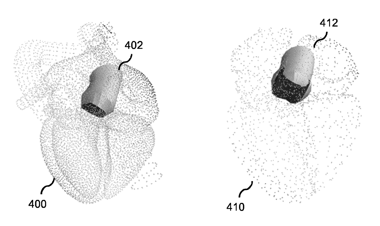
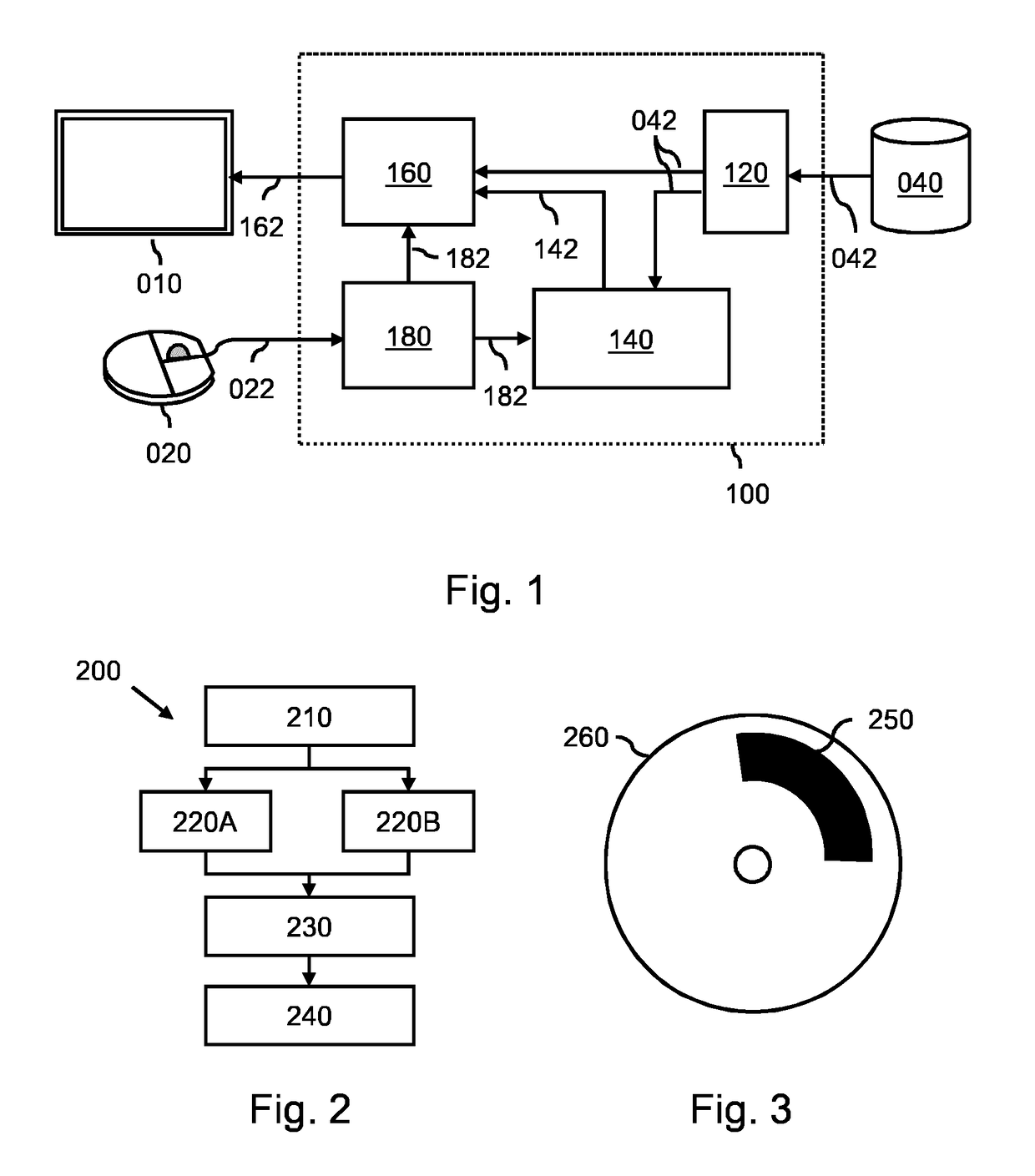
[0053]FIG. 1 shows a system 100 for fitting a deformable model to an anatomical structure in a medical image to obtain a segmentation of the anatomical structure. The system 100 comprises an image interface 120 for obtaining a first medical image of a patient and a second medical image of the patient. Both medical images show the anatomical structure and having been acquired by different medical imaging modalities or medical imaging protocols, thereby establishing a different visual representation of the anatomical structure in both medical images. FIG. 1 shows the image interface 120 obtaining the medical images in the form of image data 042 from an external database 040, such as a Picture Archiving and Communication System (PACS). As such, the image interface 120 may be constituted by a so-termed DICOM interface. However, the image interface 120 may also take any other suitable form, such as a memory or storage interface, a network interface, etc.

[0054]The system100 further compri...


PUM


Abstract

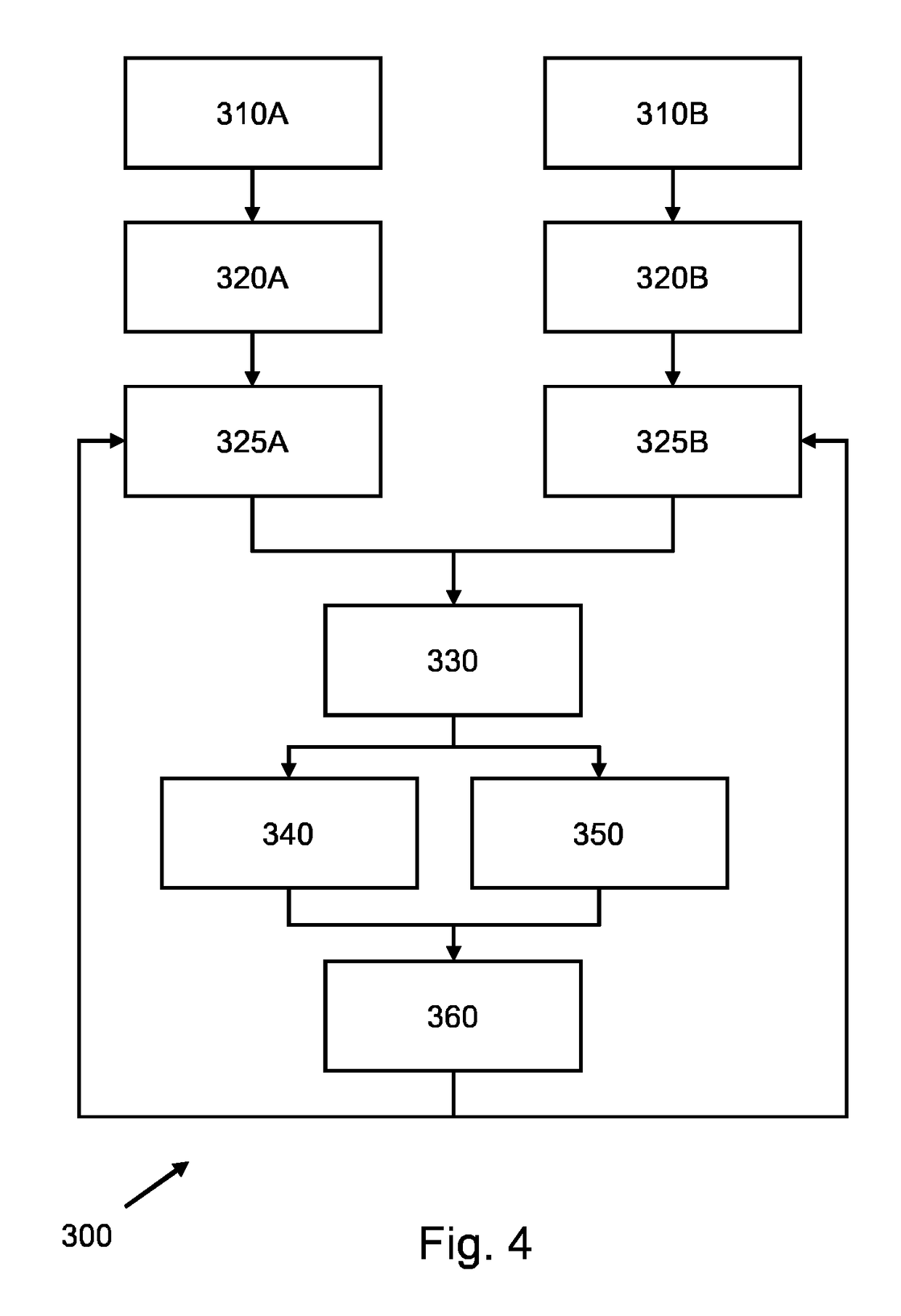
A system and method is provided which obtains different medical images (210) showing an anatomical structure of a patient and having been acquired by different medical imaging modalities and / or different medical imaging protocols. The system is configured for fitting a first deformable model to the anatomical structure in the first medical image (220A), fitting a second deformable model to the anatomical structure in the second medical image (220B), mutually aligning the first fitted model and the second fitted model (230), and subsequently fusing the first fitted model and the second fitted model to obtain a fused model (240) by augmenting the first fitted model with a part of the second fitted model which is missing in the first fitted model; or adjusting or replacing a part of the first fitted model based on a corresponding part of the second fitted model having obtained a better fit. The fused model represents a multimodal / multi-protocol segmentation of the anatomical structure, and provides a user with a more comprehensive understanding of the anatomical structure than known models.

Description

CROSS-REFERENCE TO PRIOR APPLICATIONS[0001]This application is the U.S. National Phase application under 35 U.S.C. § 371 of International Application No. PCT / EP2014 / 077660, filed on Dec. 15, 2014, which claims the benefit of European Patent Application No. 13197703.5, filed on Dec. 17, 2013. These applications are hereby incorporated by reference herein.FIELD OF THE INVENTION[0002]The invention relates to a system and method for fitting a deformable model to an anatomical structure in a medical image to obtain a segmentation of the anatomical structure. The invention further relates to a workstation and imaging apparatus comprising the system, and to a computer program product comprising instructions for causing a processor system to perform the method.BACKGROUND OF THE INVENTION[0003]Robust automatic segmentation of various anatomical structures in a medical image is a key enabler in improving clinical workflows. Here, the term segmentation refers to the identification of the anato...

Claims


Application Information

Patent Timeline
Patent Type & Authority Patents(United States)
IPC IPC(8): G06K9/00G06T7/149G06T7/00G06T7/33G06F19/00
CPCG06T7/149G06T7/344G16H30/40G06T2207/10081G06T2210/41G06T2207/20124G06T2207/20132G06T2207/30048G06T2207/10088G16H30/20
Inventor MOLLUS, SABINESAALBACH, AXELWEESE, JUERGEN
Owner KONINKLJIJKE PHILIPS NV
Learn More