
Medical navigation image output comprising virtual primary images and actual secondary images

a technology of image output and medical navigation, applied in the field of medical navigation image output, can solve the problems of affecting the freedom of movement of treatment instruments and invasive methods, and achieve the effects of improving the ergonomics of navigation methods, improving the image output of medical navigation, and optimal us

Active Publication Date: 2018-03-27
BRAINLAB
View PDF20 Cites 1 Cited by
  • Summary
  • Abstract
  • Description
  • Claims
  • Application Information

AI Technical Summary

Benefits of technology

[0005]It is therefore the object of the present invention to improve the image output in medical navigation such that available image information is optimally used. The intention is in particular also to improve the ergonomics in navigation methods and / or to minimize invasiveness.
[0008]In other words, the present invention simply reverses the previously known assembly of the image data for the image output. Navigation assistance is performed primarily on the basis of the virtual data, whereby the invention uses a technological trend, for it is possible to generate increasingly better (i.e. realistic) three-dimensional data and to process it increasingly faster and better in real time, because the corresponding data-processing hardware is becoming increasingly faster. This results in highly realistic and detailed virtual image outputs. The visual reality, i.e. actual images as perceived by the human eye, is correlated—as a second and merely augmentative form of image information—with the virtual image data and output together with it in accordance with the requirements and wishes of the user. Using such a technique in accordance with the invention, it is for example possible to more easily confirm positions of instruments when they are displayed against a less complex virtual data situation, i.e. when for example the virtual data is reduced to a particular level, such that unnecessary information is removed. Additional anatomical insights (live video) can also be provided with regard to the interactions of instruments.
[0009]Other fundamental advantages lie in the fact that it is possible to work less invasively, because the base navigation information—i.e. the virtual image data—actually already contains all the information about the “interior” of the patient, hence little direct viewing access has to be provided. In specific embodiments, the number of instruments can be reduced, and pre-operative and / or intra-operative virtual data can be updated in accordance with the interactions of the instruments.
[0013]In a preferred embodiment of the invention, the actual images are provided by a video image capture unit, in particular a camera or a camera light recorder (object lens or optical fiber end), wherein said video image capture unit is arranged on an instrument which is in particular a medical treatment instrument, specifically a pointer, a coagulation instrument, a pair of tweezers, a scalpel or a clamp. In these embodiments in particular, a reduction in the number of instruments results.
[0014]Preferably, the instrument which is navigated in accordance with the invention is specifically equipped for said navigation and can in particular be positionally detected by means of a tracking system, which can be realized by providing a tracking reference. It can also be provided as a pre-calibrated instrument, i.e. as an instrument for which the geometry is known beforehand and stored in the navigation system and which can therefore be unambiguously identified and navigated directly, without pre-calibration. It can use a plurality of instruments which are each provided with a video image capture unit, wherein the actual image data thus obtained is used to reciprocally observe or monitor the instruments and / or to produce supplementary images. Disruptive interactions can thus be avoided. Using the video image capture unit, it is also possible to detect non-visible light wavelengths which are then used for image optimization.

Problems solved by technology

In addition to the relatively marginal utilization of the virtual image material, access to the region of interest has to be uncovered when using a microscope, which makes the method more significantly invasive.
If an endoscope is used, this forms a second instrument which additionally has to be controlled and checked and which in principle can also disrupt the freedom of movement of the treatment instrument.

Method used


Image

  • Medical navigation image output comprising virtual primary images and actual secondary images
  • Medical navigation image output comprising virtual primary images and actual secondary images

Examples


Embodiment Construction

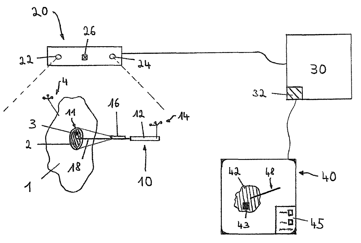
[0019]In the FIGURE, the reference sign 1 indicates—in a highly schematized form—a part of a patient's body which comprises a region of interest 2 in which a navigation-assisted treatment is to be performed. Tissue situated in the region of interest 2 has been examined beforehand using medical imaging, and a virtual data set of said tissue has been produced (for example using a magnetic resonance and / or magnetic resonance method). This tissue is indicated by hatching. An object which can be visually captured particularly easily is also imaged in the region of interest 2 and bears the reference sign 3.

[0020]A reference array 4 is situated on the part of the patient's body. This reference array 4 and a reference array on the surgical instrument 10 (likewise shown) are situated in the visual range of a tracking system 20 which positionally detects and tracks these reference arrays 4 and 14 using two cameras 22 and 24, such that where the instrument 10 and the part of the body 1 are sit...


PUM


Abstract

The invention relates to an image output method for medical navigation in which the positional relationship of an instrument with respect to a part of a patient's body is ascertained, and in which the instrument and the part of the patient's body are displayed in the correct positional relationship on an image output, wherein the display of the part of the patient's body is based on the one hand on virtual image data captured by means of a medical imaging method and on the other hand on actual images captured during navigation, wherein the virtual image data is displayed on the image output primarily and as the basis of the image, and the actual images are superimposed on it merely as an addition and secondarily. It also relates to an image generating and image output device for medical navigation.

Description

RELATED APPLICATION DATA[0001]This application claims the priority of U.S. Provisional Application No. 61 / 165,551, filed on Apr. 1, 2009, which is hereby incorporated in its entirety by reference.FIELD OF THE INVENTION[0002]The invention relates to the technical field of medical navigation image output. Medical navigation is used within the framework of image-guided surgery and assists surgeons in optimally positioning their instruments, wherein for example reference is made to patient image data acquired beforehand. As a result, the physician performing the treatment is assisted by an image output, for example a monitor, on which they can identify where their instrument or its functional portion is situated in relation to particular parts of a patient's body—regions of interest. This technology is specifically advantageous in minimally invasive types of treatment, in which instruments are for example introduced through small openings in the surface of the patient's skin and in whic...

Claims


Application Information

Patent Timeline
Patent Type & Authority Patents(United States)
IPC IPC(8): H04N7/18A61B34/20A61B90/00A61B1/06A61B1/05A61B1/04A61B5/06H04N5/225G02B21/36A61B1/07
CPCA61B90/36A61B34/20A61B1/041A61B1/042A61B1/05H04N2005/2255A61B1/07A61B5/06A61B2090/364A61B2090/365G02B21/365A61B1/0638H04N23/555
Inventor SCHMIDT, ROBERT
Owner BRAINLAB
Learn More