
Biodegradable nerve guides

a nerve guide and biodegradable technology, applied in the field of biodegradable nerve guides, can solve the problems of device failure, inflammation, growth factor inactivation, leakage of growth factors from the nerve guide, etc., and achieve the effect of reducing the burst release of protein and prolonging the releas

Active Publication Date: 2017-07-18
THE JOHN HOPKINS UNIV SCHOOL OF MEDICINE
View PDF21 Cites 9 Cited by
  • Summary
  • Abstract
  • Description
  • Claims
  • Application Information

AI Technical Summary

Benefits of technology

This approach enables controlled and sustained release of neurotrophic factors, enhancing axonal regeneration by providing a conducive biochemical environment and mechanical guidance, leading to improved nerve repair outcomes comparable to autologous nerve grafts.

Problems solved by technology

Alongside the fact that even autografts are successful only 50% of the time, problems such as donor-site morbidity and donor nerve shortage prompt the search for alternative methods for promoting peripheral nerve regeneration.
Problems with this approach include leakage of growth factors from the nerve guide and growth factor inactivation.
While continuous delivery devices such as osmotic pumps and silicone reservoirs have been used to overcome these problems, they also suffer from complications such as device failure and inflammation resulting from the non-degradable components.
Complications with these methods arise also, however, due to the fact that growth factor release cannot be controlled tightly; some growth factors may be toxic when delivered at a high local concentration.
Even further, a problem known as the “growth-factor oasis” effect has been observed in which high levels of growth factors within the nerve guide allow axons to regenerate into the nerve guide but not out, because the regenerating axons simply become fixed to the local maximum in the growth factor concentration profile.
The poor control of neurotrophic factor encapsulation and release has been the limiting factors to develop an efficient release system to fully harness the potential benefits of local sustained release of neurotrophic factors to improve PNS regeneration.
The complication is that a) loading neurotrophic factors with controlled dose and release kinetics has been difficult; b) growth factor loading involves organic solvent and harsh conditions, which will significantly decrease the bioactivities of the growth factors; c) it is difficult to adjust the total dose of growth factors and the nanofibers independently.

Method used


Image

  • Biodegradable nerve guides
  • Biodegradable nerve guides
  • Biodegradable nerve guides

Examples


example 1

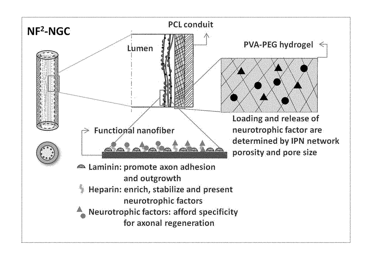
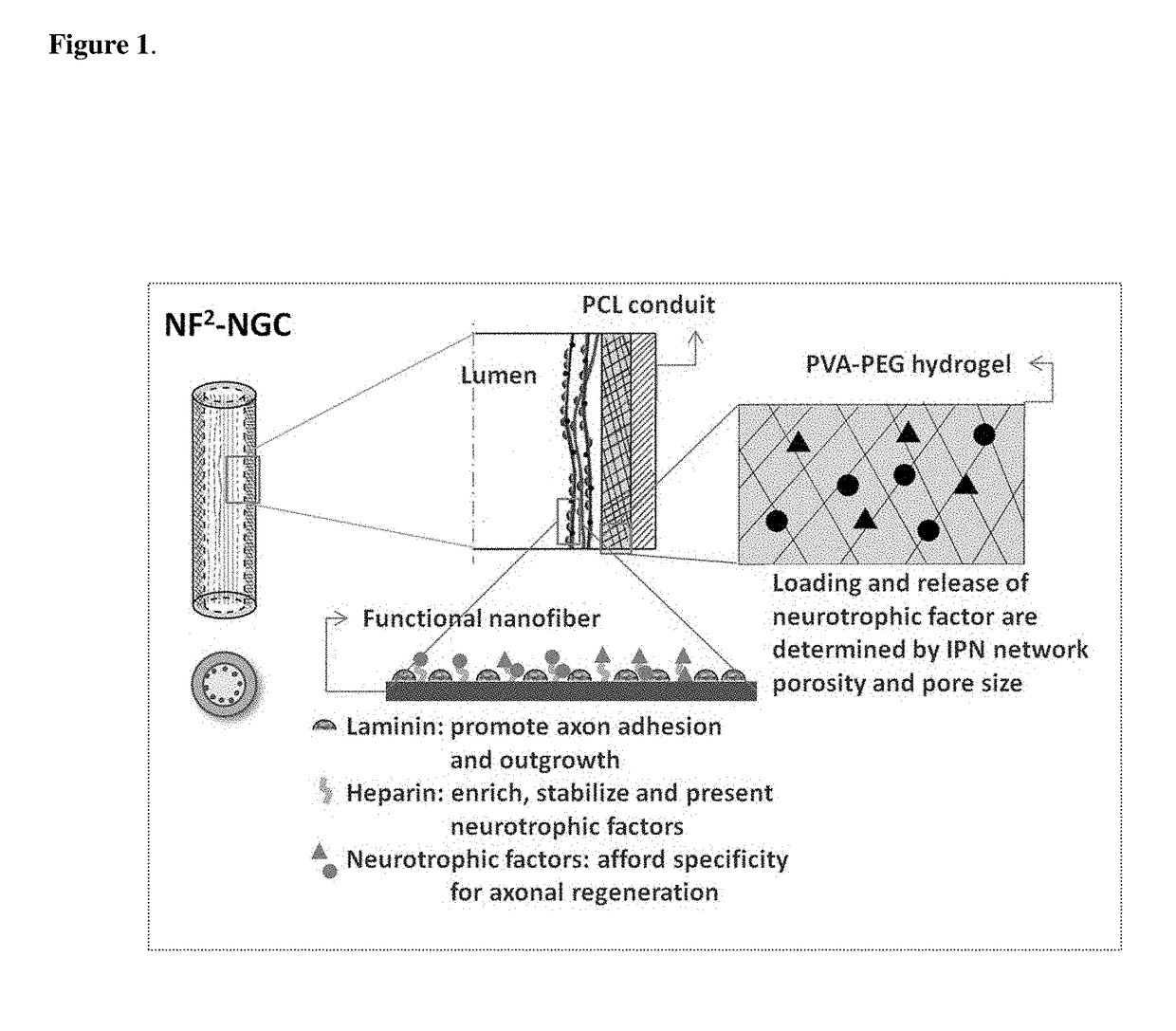
Preparation of a Nerve Guide

[0080]1) Cast a 10 cm2 film of 50:50 PCI / PCLEEP on top of a glass slide mold from a 12% wt solution in dichloromethane.[0081]2) Dry the film under vacuum over night to remove residual solvent.[0082]3) Place slide on a hot plate and heat the film to 100° C.[0083]4) Add 1 ml of 1% PVA in water on top of the film.[0084]5) Quench the film at −20° C. for 30 minutes.[0085]6) Add 500 μL of 5% PVA containing 5 μg GDNF on top of the film.[0086]7) Freeze the composite at −20° C. for 12 hours.[0087]8) Cool at 25° C. for 30 minutes.[0088]9) Freeze the composite at −20° C. for 2 hours.[0089]10) Cool at 25° C. for 30 minutes.[0090]11) Repeat steps 9 and 10 for a total of 12 cycles.[0091]12) Freeze-dry the construct.[0092]13) Place electrospun PCL nanofibers modified with laminin on top of the hydrogel layer.[0093]14) Roll the construct into a tube with inner diameter of 3 mm and the hydrogel layer on the luminal side.[0094]15) Seal the membrane with a trace quantity of...

example 2

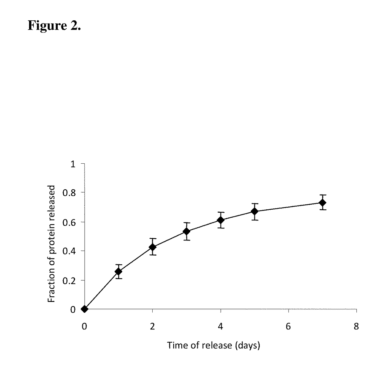
Release Profile of Model Proteins from the Nerve Guide Construct

[0096]A nerve guide is prepared as described in Example 1 with bovine serum albumin (BSA) as a model protein factor loaded in a 5% PVA-83k-99 hydrogel construct into buffered saline at 37° C., gel was frozen at −2° C. for 72 hours, followed by 4 conventional freeze-thaw cycles. The release profile of BSA is shown in FIG. 2.

example 3

Process of Preparing a Double-Layered Hydrogel Nerve Guide

[0097]1) Repeat steps 1-8 as described in Example 1 with Gel A loaded onto the PCL membrane.[0098]2) Independently prepare a 0.5 mm thick PVA hydrogel (Gel B) by replicating steps 6-11 as described in Example 1, with the sole modification of omitting the growth factor loading.[0099]3) Add 50 μL of 5% PVA solution on top of Gel A, then lay Gel 13 on top of the solution.[0100]4) Repeat steps 9 and 10 as described in Example 1 for a total of 4 cycles.[0101]5) Continue with steps 12-16 as described in Example 1.

[0102]Other modifications can be used to modify the rate of release of proteins from the hydrogel. The addition of a protein-free top layer provides an additional diffusion barrier to incorporate time-delayed protein release. Blending PVA with other water-soluble small molecular weight polymers changes the hydrogel crystallinity. Changing the number of freeze-thaw cycles, or simply incubation for prolonged periods at sub-z...


PUM


Abstract

The present invention is directed to the compositions and methods of preparing hydrogel-grafted nerve guides for peripheral nerve regeneration. Particularly, the present invention describes the nerve guides and methods for preparation of hydrogel-grafted nerve guides with encapsulated neurotrophic factors and a nanofiber mesh lining the inner surface of the guide. The present invention also provides methods for peripheral nerve repair using these hydrogel-grafted nerve guides.

Description

RELATED APPLICATIONS[0001]This application is a continuation application of U.S. Ser. No. 12 / 864,493, filed Jan. 31, 2011 (now abandoned), which is a national stage application filed under 35 U.S.C. §371 of international application no. PCT / US2009 / 000535, filed Jan. 26, 2009, which claims the benefit of priority under 35 U.S.C. §119(e) to U.S. Provisional Application No. 61 / 062,434, filed Jan. 25, 2008, each of which is incorporated herein by reference in its entirety.GOVERNMENT SUPPORT[0002]The following invention was supported at least in part by National Science Foundation DMR-0748340. Accordingly, the government has certain rights in the invention.BACKGROUND OF THE INVENTION[0003]injuries to the peripheral nervous system can result in physical discontinuity of a peripheral nerve, leaving two uncoupled nerve ends, if the gap between these ends is relatively small (<20 mm), the nerve ends are surgically sutured together after which normal regenerative processes lead to eventual...

Claims


Application Information

Patent Timeline
Patent Type & Authority Patents(United States)
IPC IPC(8): A61B17/11A61L27/38A61L27/48A61F2/02A61L27/54A61L27/56A61L27/58A61F2/00A61L27/52
CPCA61B17/1128A61F2/0063A61F2/02A61L27/383A61L27/3878A61L27/48A61L27/52A61L27/54A61L27/56A61L27/58A61L2300/414A61L2300/602A61L2300/608A61L2400/12A61L2430/32Y10T156/10Y10T156/1043
Inventor HOKE, AHMETLIM, SHAWN H.LIU, XINGYUMAO, HAI-QUAN
Owner THE JOHN HOPKINS UNIV SCHOOL OF MEDICINE
Learn More