Determination of physiological cardiac parameters as a function of the heart rate

a physiological cardiac parameter and heart rate technology, applied in the field of physiological cardiac parameters as a function of heart rate, to achieve the effect of improving the reconstruction of three-dimensional image data sets, noise effects and other artifacts in image data sets

Active Publication Date: 2016-08-30
SIEMENS HEALTHCARE GMBH
View PDF23 Cites 2 Cited by
  • Summary
  • Abstract
  • Description
  • Claims
  • Application Information

AI Technical Summary

Benefits of technology

[0014]The procedure according to one or more of the present embodiments also includes the calculation of the usual angiography image data sets (e.g., the calculation of four-dimensional coronary angiography image data sets), and additionally offers an improved determination of physiological cardiac parameters as a function of the heart rate, such that overall a combination of a kind that hitherto could only be achieved in the prior art by a plurality of image recordings using different methods is produced. The method according to one or more of the present embodiments therefore provides a reproducible, heart-rate dependent functional imaging of the heart (e.g., using an X-ray device that has a C-arm).
[0019]This involves digital subtraction angiography images, (e.g., at least images recorded after the administration of a contrast agent). The recording of the four-dimensional image data sets may take place after an administration of contrast agent. In one embodiment, a fresh administration of contrast agent may take place for each heart rate, or a plurality of heart rates may be measured after an administration of contrast agents. In this context (e.g., when cardiac perfusion is to be investigated as a physiological cardiac parameter), at least one late-enhancement image data set may be recorded for each heart rate. By appropriate timing and delayed recording, it is consequently possible to determine a “first-pass” and a “late-enhancement” cardiac perfusion as a function of the heart rate. As a result of heart rates being specifically adjusted by cardiac stimulation (e.g., pacing), these recordings are in principle repeatable and reproducible. This also allows extensive comparability of the records. Late-enhancement image data sets may be recorded (e.g., 10 to 30 minutes after first-pass-image data sets and show typical properties of scar tissue and suchlike).
[0024]In one embodiment, projection images of other cardiac phases and / or of other heart rates are taken into consideration in the reconstruction of the three-dimensional image data sets. Due to the basic comparability of the heart movements at different heart rates, projection images for other heart rates will provide information (e.g., for the same cardiac phase) that may improve the reconstruction of three-dimensional image data sets for the cardiac phases at other heart rates. By taking into account further image data available, noise effects and other artifacts in the image data sets may, for example, be reduced.
[0025]This is advantageous, for example, when the reconstruction is carried out as an iterative reconstruction, in which the projection images for other cardiac phases and / or other heart rates involve at least one boundary condition and / or the target function. Iterative reconstructions methods use a priori information in order to achieve an improved reconstruction of three-dimensional image data sets for the cardiac phases.

Problems solved by technology

However, this only allows numerical determinations of cardiac parameters for various heart rates to be carried out.

Method used


Image

  • Determination of physiological cardiac parameters as a function of the heart rate
  • Determination of physiological cardiac parameters as a function of the heart rate
  • Determination of physiological cardiac parameters as a function of the heart rate

Examples


Embodiment Construction

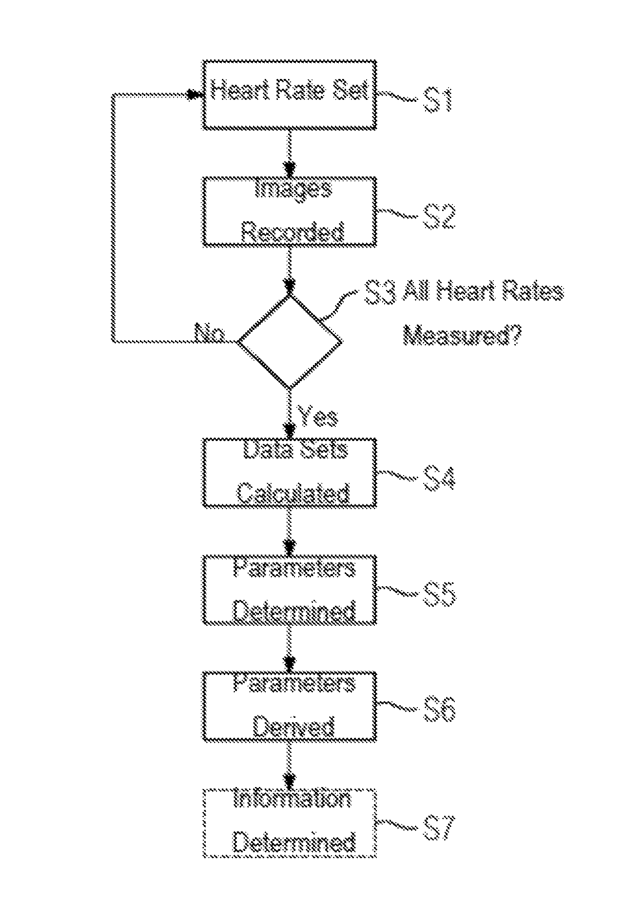
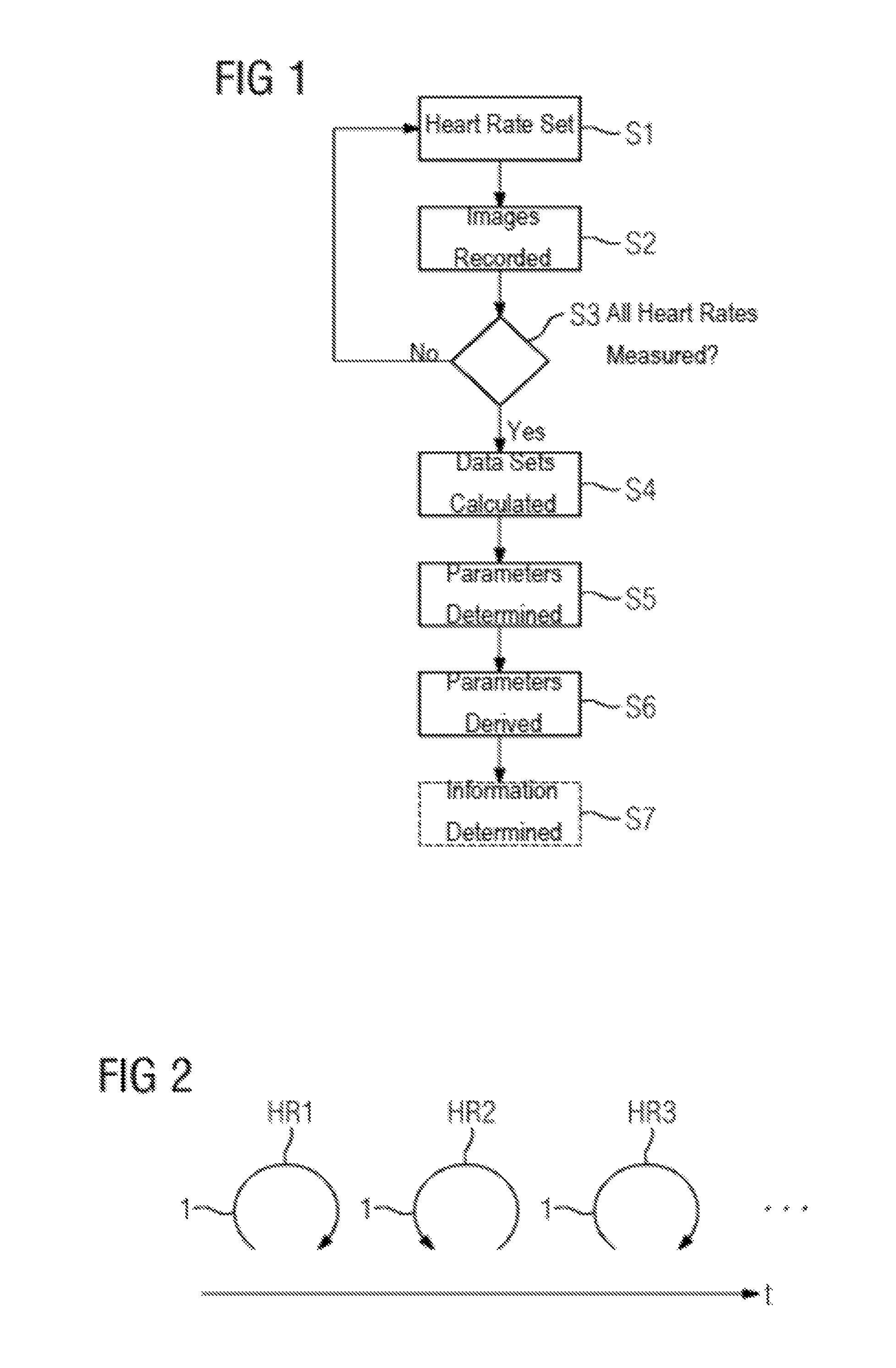
[0039]FIG. 1 shows a flow diagram of an exemplary embodiment of a method, where cardiac stimulation to adjust heart rates is also shown. Before the method is begun, a patient is positioned in an X-ray device (e.g., in an X-ray device having a C-arm, on which an X-ray emitter and an X-ray detector are arranged facing each other). Before projection images are recorded, in act S1, a first heart rate is set. The first heart rate is higher than a usual resting heart rate. In one embodiment, 80 bpm is selected as the first heart rate. The adjustment of the heart rate in the patient may be achieved, for example, by cardiac stimulation using a pacing catheter that is located in the heart, or using a cardiac pacemaker.

[0040]If the heart rate in act S1 is set at a stable rate, then in act S2, projection images for the patient are recorded from different projection directions, and such that the recording of the projection images is synchronized with the cardiac cycle. In practical terms, this ...


PUM


Abstract

A method for determining physiological cardiac parameters as a function of a heart rate is provided. For at least two heart rates adjusted by a cardiac stimulation during the recording, in each case, a four-dimensional image data set of the heart showing the entire cardiac cycle is recorded using an X-ray device. The physiological cardiac parameters are determined by evaluation of the four-dimensional image data sets.

Description

BACKGROUND[0001]The present embodiments relate to determination of physiological cardiac parameters as a function of heart rate.[0002]In the field of angiography, a number of techniques are known for obtaining information about a patient's heart. Most images (e.g., X-ray images) of the heart are taken using the technique of digital subtraction angiography (DSA). DSA involves a contrast agent being administered to the patient such that, on images of the heart, the blood vessels filled with contrast agent and the heart filled with contrast agent may be distinguished clearly. If mask images that had been taken before the contrast agent was introduced into the region to be X-rayed in a patient who, compared to the situation with the contrast images, has not been moved, are subtracted from the images taken using contrast agent (e.g., “contrast images”), all that remains, apart from noise effects, are signal components of the contrast agent. An excellent evaluation of the resulting DSA im...

Claims


Application Information

Patent Timeline
Patent Type & Authority Patents(United States)
IPC IPC(8): A61B6/00A61B6/03A61N1/365A61B5/024A61B5/00
CPCA61B6/503A61B6/481A61N1/36578A61B6/032A61B6/504A61B6/5217A61B6/4441A61B5/024A61B5/7289
Inventor KYRIAKOU, YIANNIS
Owner SIEMENS HEALTHCARE GMBH
Why Eureka
Try Eureka