
Osteosynthetic shape memory material intramedullary bone stent and method for treating a bone fracture using the same

a technology of shape memory material and bone stent, which is applied in the field of osteosynthetic shape memory material intramedullary bone stent and the same treatment method, can solve the problems of large number of fractures that require surgical intervention, lack of proper bone alignment, and impaired function, and achieve the effect of reducing fractures

Active Publication Date: 2016-03-15
ARTHREX
View PDF27 Cites 17 Cited by
  • Summary
  • Abstract
  • Description
  • Claims
  • Application Information

AI Technical Summary

Benefits of technology

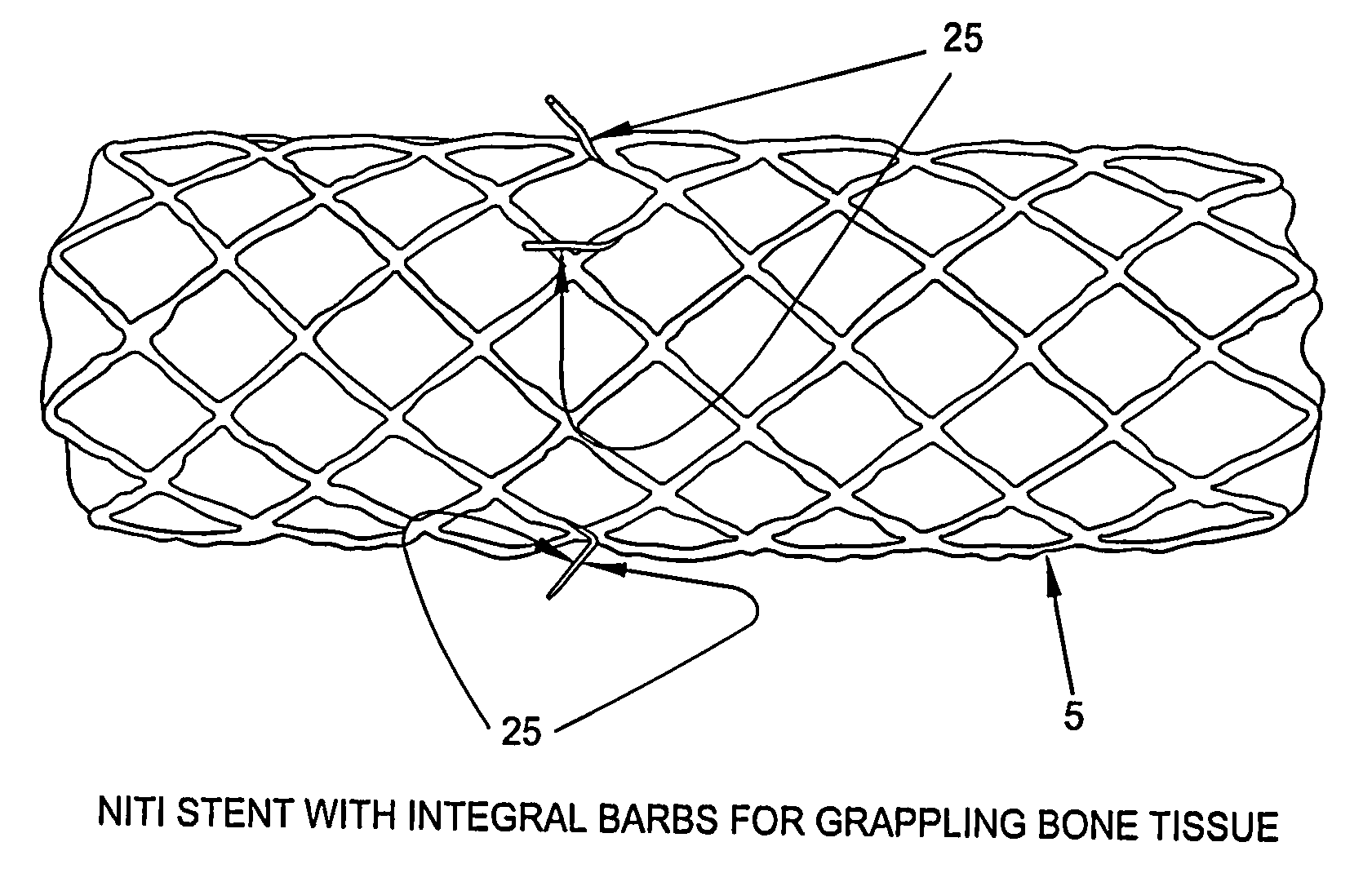
[0031]In accordance with the present invention, there are now disclosed novel intramedullary bone stents which can be used as a means of fracture fixation. Intramedullary bone stents can be made of Shape Memory Alloys (e.g., Nitinol), or other alloys such as Stainless Steel. The intramedullary bone stent is placed into the intramedullary canal, bridging the fracture. As the intramedullary bone stent expands radially, applying hoop stress to the bone, the intramedullary bone stent longitudinally contracts, reducing the fracture and rigidly holding the bone in the correct position. An intramedullary bone stent can be superior to current fixation methods because the intramedullary bone stent can have a modulus of elasticity closer to that of bone, does not create regions of stress shielding, and does not impair blood flow at the fracture site. Additionally, intramedullary bone stents may be beneficial in treating pediatric patients with open growth plates, where nails, rods, and screws may interfere with bone development at the growth plate.

Problems solved by technology

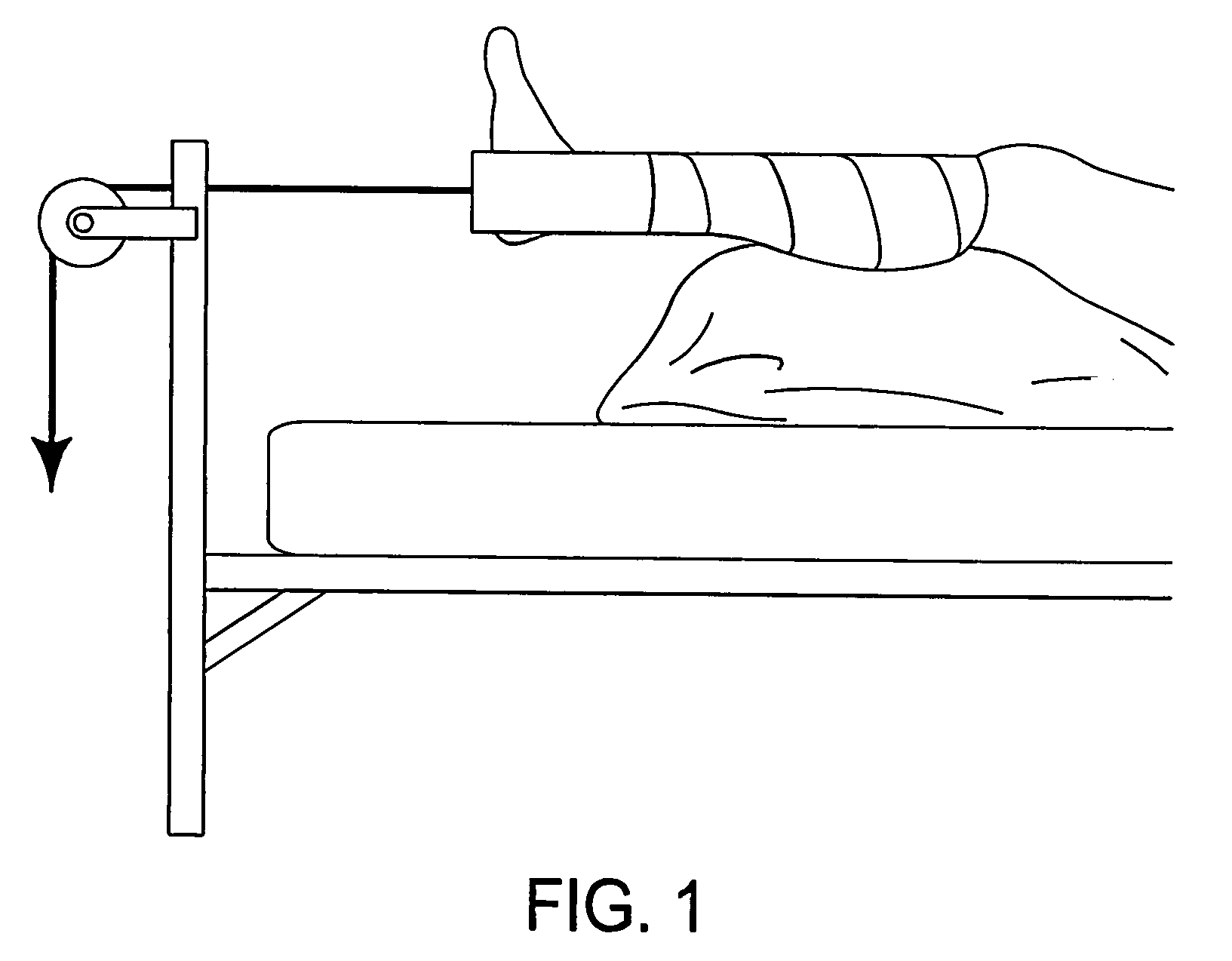
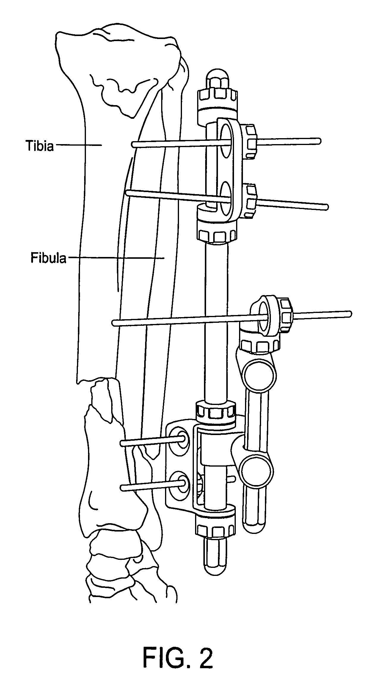
While many fractures are treated non-operatively, a large number of fractures require surgical intervention.
If a fracture that requires operative treatment is not treated, nature tries to stabilize the mobile fragments by pain-induced contraction of the surrounding muscles, which may lead to bone shortening.
The end result of this process frequently is the lack of proper bone alignment and impaired function.
Bridging of the fracture with a stiff splint reduces mobility of the fracture fragments and allows minimal displacement under functional load.
This is important because unevenness of the joint surface and presence of callus formation at the articular surface lead to patient discomfort and often development of early and progressive osteoarthritis.
However, in some cases, over time, the stainless steel and titanium fixation devices do not maintain compression across the fracture fragments.
Additionally, as the necrotic surfaces of the fracture are resorbed, a non-load bearing gap develops between the fragments, thereby decreasing compression and increasing the risk of interfragmentary motion and scar tissue formation.
Loss of compression is contrary to the objectives of fracture fixation in general.
The unmineralized scar tissue does not support load and may lead to loss of skeletal function.
One of the potential complications with cerclage wires is interruption of the periosteal blood supply with subsequent osteonecrosis or fracture nonunion.
Non-self-tapping screws are easier to insert and remove, but they are not the best choice for fixing fractures in regions with a thin cortex.
A major complication with plating is the potential compromising of cortical blood supply because of the large contact area between the plate and the underlying cortex.
Intramedullary nails provide excellent stability against bending forces, but they do not control rotation and compressive forces.
The major disadvantage associated with flexible rods is the frequent need for additional external stabilization such as a plaster cast.

Method used


Image

  • Osteosynthetic shape memory material intramedullary bone stent and method for treating a bone fracture using the same
  • Osteosynthetic shape memory material intramedullary bone stent and method for treating a bone fracture using the same
  • Osteosynthetic shape memory material intramedullary bone stent and method for treating a bone fracture using the same

Examples


Embodiment Construction

[0063]Novel Intramedullary Bone Stents: Intravascular stents are commonly used in arteries to hold the artery open, e.g., to treat stenosis caused by a buildup of plaque on the walls of the artery. Thus, these intravascular stents are designed to maintain patency of the blood vessel. Intravascular stents are inserted into the artery in a crimped state and expanded to a larger diameter. See, for example, FIG. 10, which shows expanding stents used in blood vessels. To date, stents have not been used in orthopedic applications to reduce a fracture site.

[0064]In accordance with the present invention, and looking now at FIG. 11, there is shown an intramedullary bone stent 5. Intramedullary bone stent 5 is placed into the intramedullary canal 10 of a bone 15, bridging the fracture 20. As the intramedullary bone stent 5 expands, applying hoop stress to the bone 15, the intramedullary bone stent 5 shortens, reducing the fracture 20 and rigidly holding the bone 15 in the correct position. In...


PUM


Abstract

A method for providing stabilization and compression of a bone fracture, the method comprising: providing an intramedullary prosthesis sized for insertion into the intramedullary canal of a bone and having a distal end, a proximal end and a porous structure therebetween, wherein (i) in a first state, the prosthesis is longitudinally expanded and radially contracted, and (ii) in a second state, the prosthesis is longitudinally contracted and radially expanded, conforms to the shape of the adjacent bone and exerts forces on the adjacent bone; and positioning the intramedullary prosthesis within the intramedullary canal of a bone so that the intramedullary prosthesis spans the bone fracture, whereby to provide stabilization and compression of the bone fracture.

Description

REFERENCE TO PENDING PRIOR PATENT APPLICATIONS[0001]This patent application claims benefit of:[0002](i) pending prior U.S. Provisional Patent Application Ser. No. 61 / 537,766, filed Sep. 22, 2011 by Matthew Fonte for OSTEOSYNTHETIC SHAPE MEMORY MATERIAL INTRAMEDULLARY BONE STENTS AND METHODS FOR TREATING BONE FRACTURES USING THE SAME; and[0003](ii) pending prior U.S. Provisional Patent Application Ser. No. 61 / 570,091, filed Dec. 13, 2011 by Matthew Fonte for OSTEOSYNTHETIC SHAPE MEMORY MATERIAL INTRAMEDULLARY BONE STENTS AND METHODS FOR TREATING BONE FRACTURES USING THE SAME.[0004]The two (2) above-identified patent applications are hereby incorporated herein by reference.BACKGROUND OF THE INVENTION[0005]Introduction: Osteosynthesis is the reduction and fixation of a bone fracture with an implantable device. The device is usually made of metal, either titanium or stainless steel, and may include plates, pins, rods, screws, and staples. It is a surgical procedure with an open or percu...

Claims


Application Information

Patent Timeline
Patent Type & Authority Patents(United States)
IPC IPC(8): A61B17/56A61B17/72A61B17/00
CPCA61B17/72A61B17/7216A61B17/7225A61B2017/00867A61B17/7208A61B17/7266A61B2017/00862
Inventor FONTE, MATTHEW
Owner ARTHREX
Learn More