Serological screening for HHV-8 infection using antigen mixtures
a technology of antigen mixture and serological screening, which is applied in the direction of instruments, dsdna viruses, peptides, etc., can solve the problems of limited success and limit the sensitivity
- Summary
- Abstract
- Description
- Claims
- Application Information
AI Technical Summary
Problems solved by technology
Method used
Image
Examples
example 1
Material and Methods
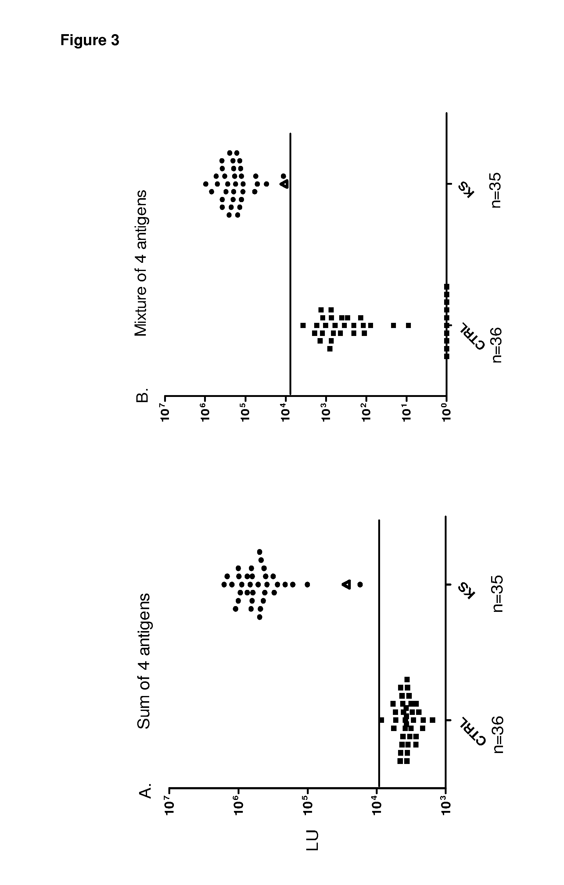
[0075]Patient sera for antibody identification. Sera for were obtained from patients or volunteers under institutional review board-approved protocols at the Clinical Center, NIH and at Georgetown University. In the initial pilot set, 6 colon cancer, 15 KS, and one HIV positive sera samples were analyzed. Subsequent analysis of the single HIV positive sample revealed that is was highly positive by both ELISA and LIPS for anti-HHV-8 antibodies and, for simplicity, it was designated as a KS sample making “16” KS samples. The training (n=83 blinded sera) and validation (n=71) sera sample sets were provided as coded samples for both LIPS and ELISA assays and the code was broken only after titers were established and categorization of HHV-8 infection status had been made. The training set consisted of 39 KS samples, while validation set contained 34 KS samples. One volunteer sample in each of the training and validation sets were also found to be HHV-8 positive by bot...
example 2
Patient Antibody Responses to Known HHV-8 Antigens
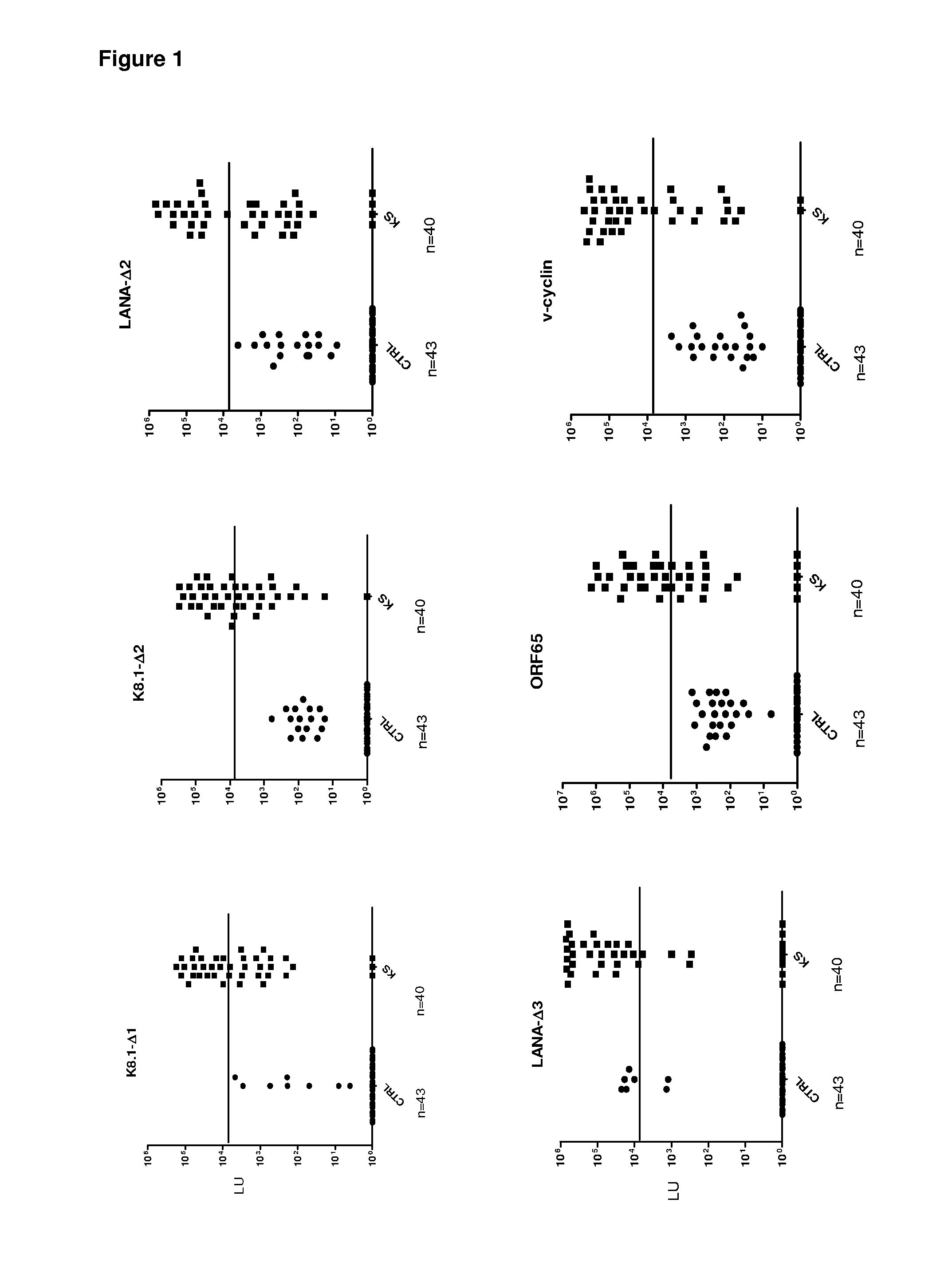
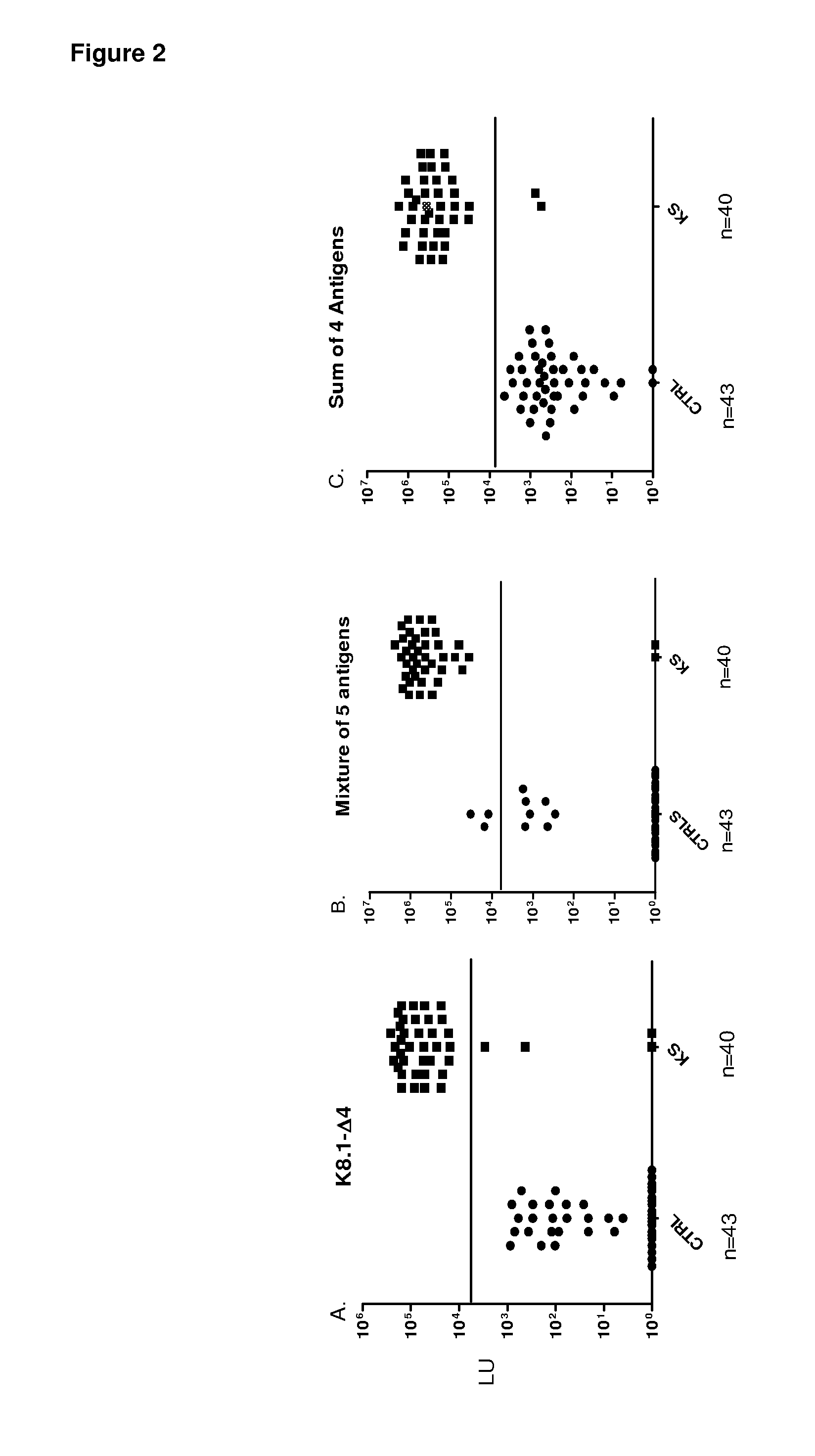
[0082]Evaluation of the utility of LIPS in HHV-8 diagnosis began by screening sera samples from 16 KS and 6 control patients for antibodies against known HHV-8 antigens, including LANA, ORF65 and K8.1. Because of the small pilot set, the discriminatory potential of each of these HHV-8 antibody tests was evaluated by a variety of statistical and diagnostic methodology including receiver operator characteristics (ROC), area under the curve (AUC), Whitney-Mann U tests and using sensitivity and specificity calculations derived from the mean plus 5 SD of the control samples (Table 1).
[0083]In the case of LANA, we generated three different protein fragments designated LANA-Δ1, LANA-Δ2, and LANA-Δ3, respectively. Using the N-terminal LANA-Δ1 fragment, only one of the 16 KS sera showed positive results by the cut-off compared to the controls (Table 1). In contrast, LANA-Δ2, representing the central region of LANA, detected positive antibody ...
example 3
Identification of v-cyclin as a New Informative HHV-8 Antigen
[0085]To identify potentially new HHV-8 antigens, the pilot sera set was screened with a panel of 14 different HHV-8 fusion proteins including v-cyclin, vFLIP / ORF71 / K13, v-BCL2 / ORF16,vIL-6 / K2, v-GPCR / Orf74, ORF48, ORF52, Kaposin / K12, ORF57, vIRF-1 / ORF K9, vIRF-3 / LANA2, ORF45, ORF47 and MCP-1 Most of these HHV-8 proteins showed weak or non-existent antibody signals with the KS sera (Table 1 and data not shown). For example, ORF52 showed weak positive signals in only 3 of the 16 KS sera (Table 1), while ORF74 showed no signal in any of the sera tested (data not shown). However, the latent HHV-8 protein, v-cyclin, displayed positive results in 12 of 16 KS samples (Table 1). The geometric mean titer of the anti-v-cyclin antibody in the 16 KS samples was 14,281 LU (95% CI, 1,318-154,759 LU), 1000 fold higher than the geometric mean titer of 15 LU (95% confidence interval [CI], 0.65-56) in the controls (Mann Whitney U test, P<0....
PUM
| Property | Measurement | Unit |
|---|---|---|
| length | aaaaa | aaaaa |
| antibody titers | aaaaa | aaaaa |
| antibody titer | aaaaa | aaaaa |
Abstract
Description
Claims
Application Information
 - R&D
- Intellectual Property
- Life Sciences
- Materials
- Tech Scout
- Unparalleled Data Quality
- Higher Quality Content
- 60% Fewer Hallucinations
Browse by: Latest US Patents, China's latest patents, Technical Efficacy Thesaurus, Application Domain, Technology Topic, Popular Technical Reports.
© 2025 PatSnap. All rights reserved.Legal|Privacy policy|Modern Slavery Act Transparency Statement|Sitemap|About US| Contact US: help@patsnap.com


