
Methods of assaying sensitivity of cancer stem cells to therapeutic modalities

a cancer stem cell and sensitivity technology, applied in the field of single cancer stem cell proliferative ability measurement, can solve the problems of low plating efficiency, inconvenient identification of stem cells, and inability to predict individual patient outcomes, so as to achieve the effect of csc growth and maintenance of stem cell properties

Active Publication Date: 2015-01-20
THE RES FOUND OF STATE UNIV OF NEW YORK
View PDF2 Cites 0 Cited by
  • Summary
  • Abstract
  • Description
  • Claims
  • Application Information

AI Technical Summary

Benefits of technology

The present invention is a method for creating a safe and suitable environment for stem cells to grow and maintain their properties. This is done by combining tumor cells with fibroblasts to form a hybrid spheroid, which allows for the growth of cancer stem cells. The technical effect is the creation of a safe and suitable microenvironment for stem cells to grow and maintain their properties, potentially leading to new therapies for cancer treatment.

Problems solved by technology

However, stem cell identification is still problematic as the marker combinations which purportedly identify prospective hematopoietic stem cells (the best characterized) label a larger fraction (˜2-fold) of cells than that which can repopulate the bone marrow (the functional endpoint) [Yilmaz O. H. et al., Blood 2006, 107(3): 924-930; Kiel M. J. et al., Cell 2005, 121: 1109-1121]; for breast, the fraction is 100-fold [Al-Hajj M. et al., Proc Natl Acad Sci USA 2003, 100: 3983-3988].
The problem with this approach is that these markers do not identify such cells since: 1) the markers do not specify any function of the CSCs, they only help to enrich for subpopulations containing them; 2) cells possessing the appropriate markers (and lacking the inappropriate ones) are only enriched for CSCs and only a fraction of them can initiate a tumor; and 3) some cells lacking the appropriate markers supposedly defining CSCs are also able to initiate a tumor.
Thus, the prior art methods normally enrich, rather than identifying, cancer or normal stem cells.
The previous methods, which employ monolayer and agar colony formation did not prevent stromal fibroblast outgrowth from producing artifactual colonies so that the cancer cell (not cancer stem cell) survival curves obtained by those methods were not useful or meaningless for predicting individual patient outcomes.
The low plating efficiency (PE=10−5) made it difficult to claim that the results were representative of the tumor in situ.
Moreover, like all single-cell plating assays, there is a fundamental problem that soft-agar assays lack physiological relevance to in situ tumors, because they lack the three dimensional (3D) cell-cell contact seen in tumors, and the lack of a stem cell niche may be critical [Schofield, R., Blood Cells 1978, 4: 7-25; Stausbol-Gron B. et al., Radiother Oncol 1995, 37(2): 87-99; Stausbol-Gron B. et al.

Method used


Image

  • Methods of assaying sensitivity of cancer stem cells to therapeutic modalities
  • Methods of assaying sensitivity of cancer stem cells to therapeutic modalities
  • Methods of assaying sensitivity of cancer stem cells to therapeutic modalities

Examples


example 1

Research Methods

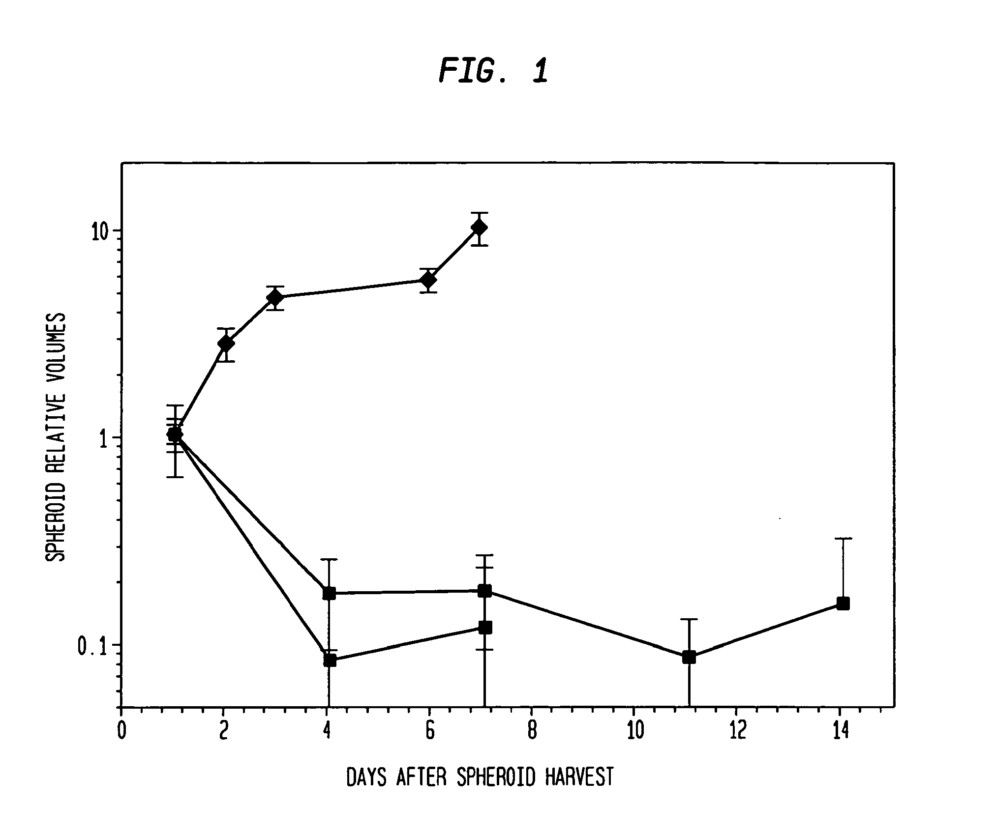
[0048]Types of spheroids: Two types of spheroids are used in this study: (1) Pure fibroblast spheroids, in which all the cells are fibroblasts; (2) Hybrid spheroids, in which two cell types are mixed (primary breast cancers, mixed with fibroblasts), one type being the test cells whose growth and viability are being measured (0.5% to 40% of the initial spheroid cells), while the other type consists of nonclonogenic fibroblast feeder cells (99.5% to 60% of the initial spheroid cells; rendered such by culture condition).

[0049]Cell lines used: Two cell types can be used: human diploid fibroblasts (either AG1522 early passage male or RMF-EG hTERT immortalized GFP labeled female) retrieved from liquid nitrogen storage shortly before use, and cells obtained directly from breast cancer surgical samples.

[0050]Tumors to be studied: Tumors tissues are obtained from patients to be analyzed. For the examples provided below, Dr. Schwartzman, Chief of General Surgery in the Surgery...

example 2

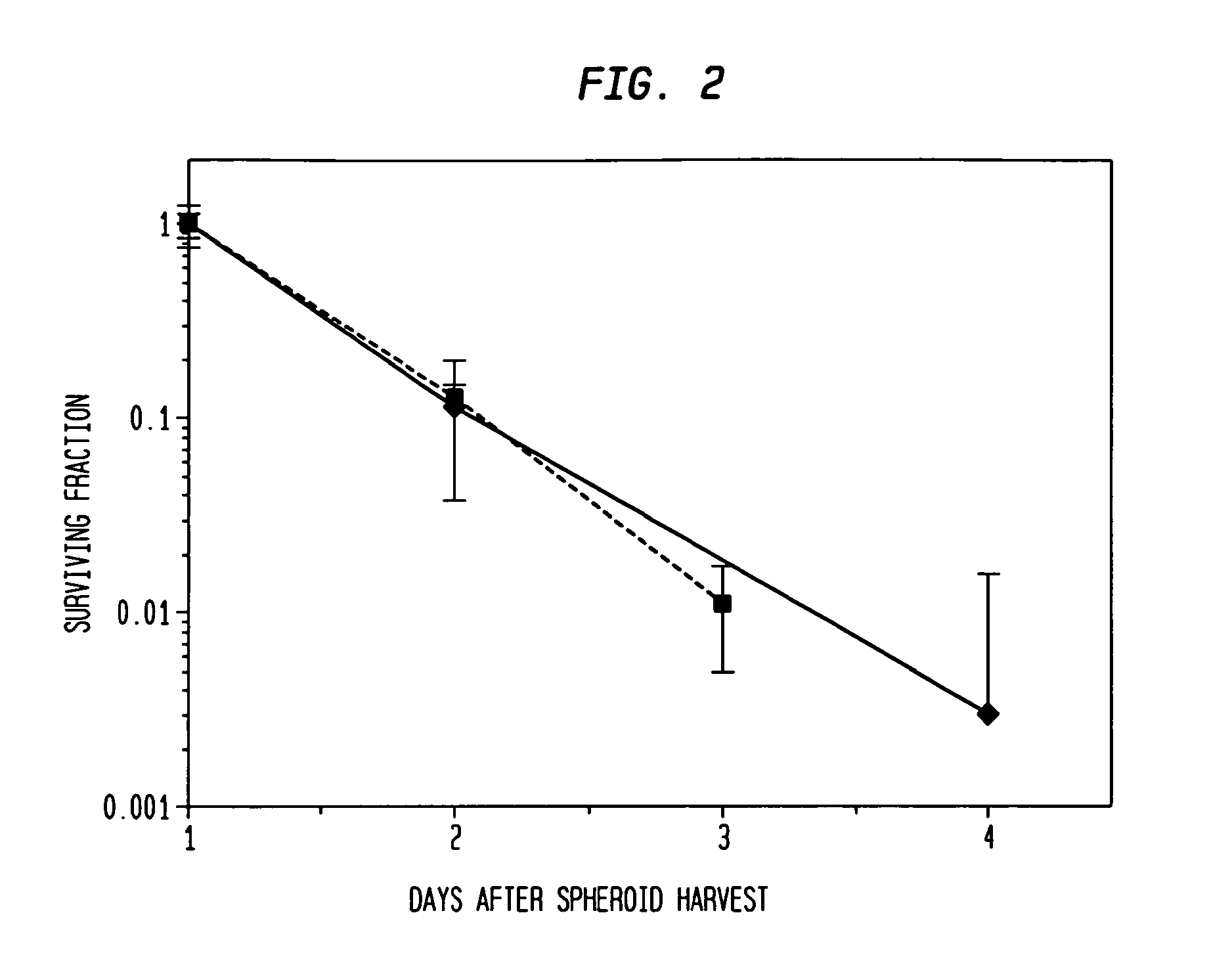
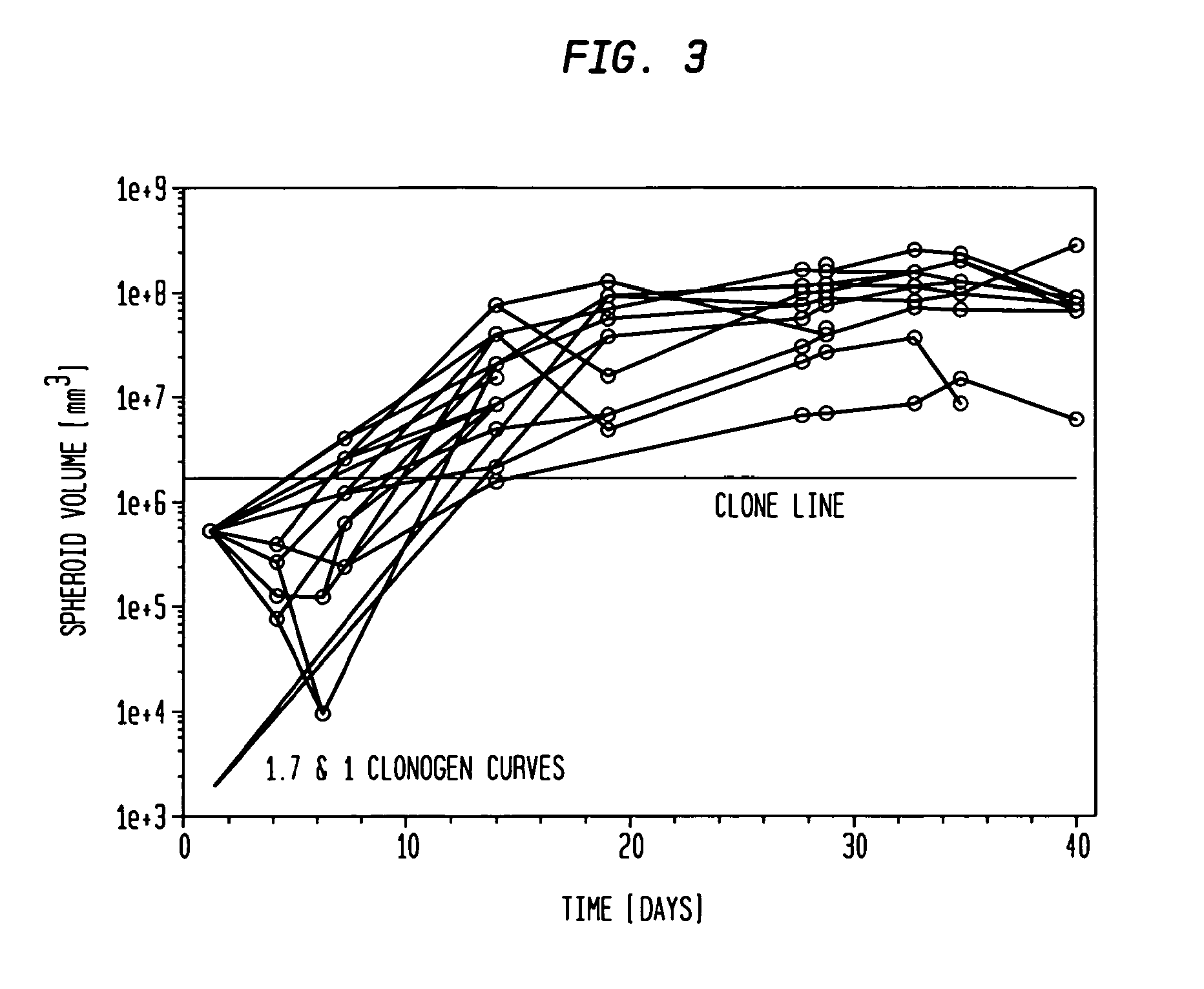
[0070]The hybrid spheroid (hs) assay: To alleviate the problems in the prior art methods, an in vivo-like system, the hs assay suitable for testing primary tumor cells has been developed [Djordjevic B. et al., Acta Oncologica, 2006, 45, 412-420; Djordjevic B. et al., Radiat. Res. 53rd Annual Meeting Program Book p 99 (PS179), Nov. 6-9, 2006, Philadelphia, Pa.; and Djordjevic B. et al., Int J Radiat Oncol Biol Phys 1992; 24(3): 511-518; Lange C. S. et al., Int J Radiat Oncol Biol Phys 1992, 24(3): 511-518; Djordjevic, B. et al., Cancer Invest 1991, 9(5): 505-512; Djordjevic B. et al., Cancer Invest. 1993, 11(3): 291-298; Djordjevic B. et al., Acta Oncologica 1998, 37: 735-739; Djordjevic B. et al., Radiat Res 1998, 150:275-282; Djordjevic B. et al., Indian J Exper Biol 2004, 42: 443-447] (see below). The system exhibits a much higher PE, ˜0.5-2% [Djordjevic B. et al., Acta Oncologica, 2006, 45, 412-420], with almost all samples producing sufficient colonies for assay [Djordjevic B. e...


PUM

PropertyMeasurementUnit
diameteraaaaaaaaaa
timeaaaaaaaaaa
doubling timeaaaaaaaaaa

Abstract

The present invention is directed to methods of measuring the proliferative ability of individual patient cancer stem cells. The present invention provides a method for treating a cancer patient according to an assay of the individual patient's tumor's cancer stem cell sensitivity, by measuring the proliferative ability of cancer stem cells from the patient. By the methods of the present invention it is possible to treat individual cancer stem cells presented in tumor cells. Methods of detecting and enumerating cancer stem cells in hybrid spheroids comprised of fibroblasts and tumor cells are also provided by the present invention. The present invention also contemplates a method for drug and other treatment development, wherein the effects of a drug or combination of drugs or other treatments are determined on the individual patient's cancer stem cells.

Description

CROSS REFERENCE TO RELATED APPLICATIONS[0001]This application is a 371 of PCT / US08 / 04247 filed on Mar. 31, 2008, which claims benefit of U.S. Provisional Applications 61 / 036,739 filed Mar. 14, 2008 and 60 / 920,922 filed Mar. 30, 2007, the contents of all of which are incorporated herein by reference.FIELD OF THE INVENTION[0002]The present invention is related to methods of measuring the proliferative ability of single cancer stem cells. Specifically, the present invention is related to methods for measuring the proliferative ability of an individual patient's cancer stem cells by obtaining a single cell suspension from the patient's tumor, engulfing them in a manner that yields an average of a single cancer stem cell in a spheroid of fibroblasts, and assaying the proliferative ability of the cancer stem cells.BACKGROUND OF THE INVENTION[0003]It is well established that many normal tissues are maintained and regenerated by a small subset of pluri- (or toti-) potential stem cells (SC) ...

Claims


Application Information

Patent Timeline
Patent Type & Authority Patents(United States)
IPC IPC(8): G01N33/50G01N33/574C12N5/00
CPCC12N2503/02C12N5/0062C12N2502/1323G01N33/5011G01N2800/52G01N33/574
Inventor LANGE, CHRISTOPHER S.DJORDJEVIC, BOZIDARROTMAN, MARVIN Z.
Owner THE RES FOUND OF STATE UNIV OF NEW YORK
Learn More