System and method for computer aided septal defect diagnosis and surgery framework

a technology of computer aided septal defect and diagnostic framework, which is applied in the field of system and method for improving the workflow of medical imaging system, can solve the problems of time-consuming manual search, increased labor intensity, and increased labor intensity

Active Publication Date: 2014-04-15
GENERAL ELECTRIC CO
View PDF16 Cites 4 Cited by
  • Summary
  • Abstract
  • Description
  • Claims
  • Application Information

AI Technical Summary

Problems solved by technology

For instance, internal defects in an object may be detected.
A time-consuming manual search is then performed to locate the septal defects.
When an echocardiogram is used, the time—consumption is even greater since it takes a relatively long time to locate the defect positions, which adds extra pain to the patient, especially after surgery.

Method used


Image

  • System and method for computer aided septal defect diagnosis and surgery framework
  • System and method for computer aided septal defect diagnosis and surgery framework
  • System and method for computer aided septal defect diagnosis and surgery framework

Examples


Embodiment Construction

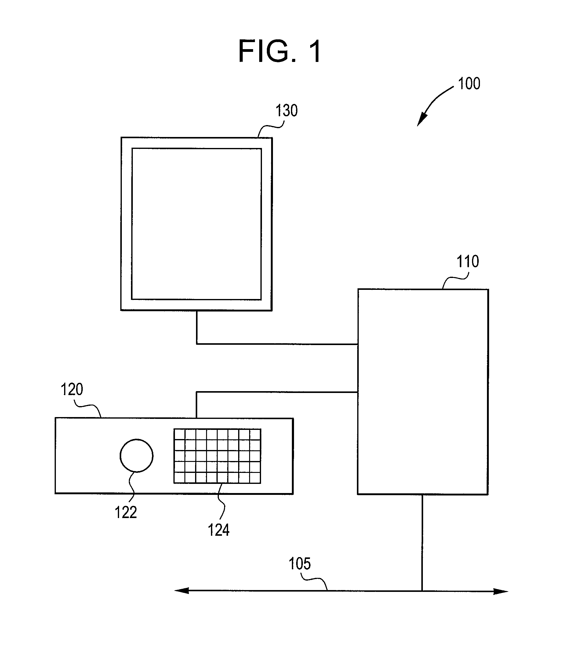
[0019]FIG. 1 illustrates a system 100 for manipulating and displaying medical images. The system 100 includes a computer unit 110. The computer unit 110 may be any equipment or software that permits electronic medical images, such as x-rays, ultrasound, CT, MRI, gated MRI, EBT, MR, or nuclear medicine for example, to be electronically acquired, stored, or transmitted for viewing and operation. The computer unit 110 may receive input from a user. The computer unit 110 may be connected to other devices as part of an electronic network. In FIG. 1, the connection to the network is represented by line 105. The computer unit 110 may be connected to network 105 physically, by a wire, or through a wireless medium. In an embodiment, the computer unit 110 may be, or may be part of, a picture archival communication system (PACS).

[0020]The system 100 also includes an input unit 120. The input unit 120 may be a console having a track ball 122 and keyboard 124. Other input devices may be used to ...


PUM


Abstract

Certain embodiments of the present invention provide systems and methods for detecting septal defects. In an embodiment, the method may include acquiring gated MRI data and heart atlas data. A registration of the gated MRI data and the heart atlas data may be performed as well as a flow-balance measurement. An atrial septal detection and ventricular septal detection may be performed, as well as a septal measurement. A diagnostic report may be generated detailing the location and properties of the septal defects. A physician may utilize the diagnostic report for surgery planning. After surgery, the diagnostic report may be compared with a post-surgery diagnostic report to determine the success of the surgery.

Description

RELATED APPLICATIONS[0001][Not Applicable]FEDERALLY SPONSORED RESEARCH OR DEVELOPMENT[0002][Not Applicable]MICROFICHE / COPYRIGHT REFERENCE[0003][Not Applicable]BACKGROUND OF THE INVENTION[0004]The present invention generally relates to a system and method for improved workflow of a medical imaging system. Particularly, the present invention relates to a more efficient system and method for diagnosing, planning surgery, and post surgery follow up for septal defects.[0005]Medical diagnostic imaging systems encompass a variety of imaging modalities, such as x-ray systems, computerized tomography (CT) systems, ultrasound systems, electron beam tomography (EBT) systems, magnetic resonance (MR) systems, and the like. Medical diagnostic imaging systems generate images of an object, such as a patient, for example, through exposure to an energy source, such as x-rays passing through a patient, for example. The generated images may be used for many purposes. For instance, internal defects in a...

Claims


Application Information

Patent Timeline
Patent Type & Authority Patents(United States)
IPC IPC(8): A61B5/05
CPCA61B5/7289A61B5/055
Inventor LI, SHOUGILL, SUKHDEEPMAHESH, PRAKASHROEDER, DAVE
Owner GENERAL ELECTRIC CO
Why Eureka
Try Eureka