
Non-invasive in-vivo imaging of mechanoreceptors in skin using confocal microscopy

a mechanoreceptor and in vivo imaging technology, applied in the field of in vivo imaging of mechanoreceptors in skin using confocal microscopy, can solve the problems of inability to evaluate the sensory nerve terminals in the skin, inability to use serial monitoring, non-invasive human in-vivo imaging approaches, etc., and achieve the effect of wide applicability for identification and monitoring

Active Publication Date: 2012-09-04
CALIBER IMAGING & DIAGNOSTICS +1
View PDF40 Cites 4 Cited by
  • Summary
  • Abstract
  • Description
  • Claims
  • Application Information

AI Technical Summary

Benefits of technology

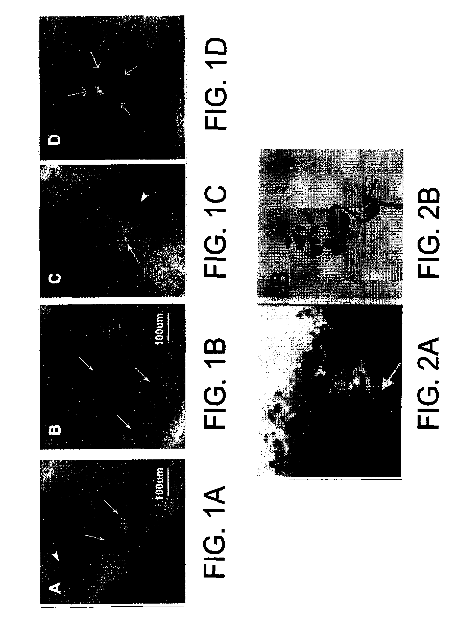
The invention is about a method to visualize and quantitate mechanoreceptors (MCs) in human or animal skin using in-vivo RCM (Removing the Catheter with Minimal Trauma). This technique can be used to diagnose and monitor peripheral nervous system disorders, such as diabetic and idiopathic SN (Sensory Neuropathy). The method is non-invasive, painless, and can be used to screen for risk factors and assess the efficacy of therapies. The invention has wide applicability and can be extended to other peripheral neuropathies or even broader peripheral nervous system disorders.

Problems solved by technology

However, biopsy assessment of MC density has not been widely used, in part as glabrous skin biopsies are more painful and invasive than biopsies of hairy skin used for ENFD counts, require complex processing, and are not suitable for serial monitoring of SN.
Non-invasive, human in-vivo imaging approaches, for evaluation of sensory nerve terminals in the skin are not available.
However, in-vivo RCM of skin has not yet been applied to measurements of MC density or to diagnostic techniques relating to neurological disorders including SN.

Method used


Image

  • Non-invasive in-vivo imaging of mechanoreceptors in skin using confocal microscopy
  • Non-invasive in-vivo imaging of mechanoreceptors in skin using confocal microscopy
  • Non-invasive in-vivo imaging of mechanoreceptors in skin using confocal microscopy

Examples


Embodiment Construction

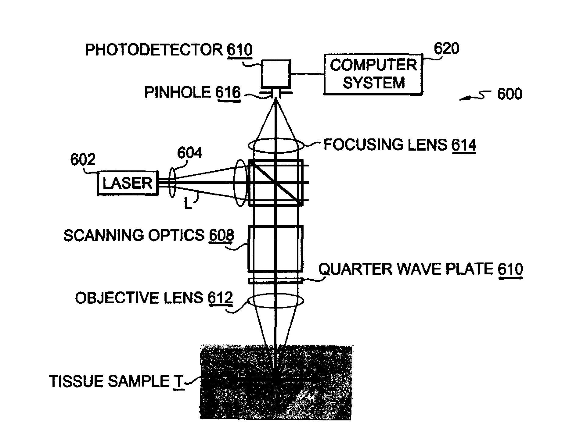
[0030]A preferred embodiment of the present invention will be set forth in detail with reference to the drawings, in which like reference numerals refer to like elements or steps throughout.

[0031]Fifteen adult subjects were recruited to participate in this pilot feasibility study, under a Rochester Subjects Review Board Approved Protocol. Enrolled subjects included 10 healthy adult controls, with no risk factors, symptoms or clinical evidence of a polyneuropathy or mononeuropathy, and 5 subjects with SN (HIV infection 3, Diabetes Mellitus 1, sensory neuronopathy with SLE 1) diagnosed through the neurology clinics at University of Rochester.

[0032]In-Vivo Confocal Microscopy Procedure.

[0033]A trained technician performed in-vivo RCM within a standardized 3×3 mm skin area over the mid-point of the volar aspect of the distal phalanx of digit V and over the mid point of the thenar eminence of the non-dominant hand in each subject. An in-vivo RCM, (VivaScope 1500, Lucid, Inc, Rochester, N...


PUM

PropertyMeasurementUnit
sizeaaaaaaaaaa
thicknessaaaaaaaaaa
depthaaaaaaaaaa

Abstract

Meissner's Corpuscles (MCs) are touch-pressure sensation receptors in glabrous skin. They are imaged by reflectance confocal microscopy to provide a non-invasive, in vivo quantification of their density or size to allow screening for, diagnosis or monitoring of sensory neuropathy and other peripheral nervous system disorders related to diabetes, HIV, or other conditions.

Description

REFERENCE TO RELATED APPLICATION[0001]The present application claims the benefit of U.S. Provisional Patent Application No. 60 / 833,240, filed Jul. 26, 2006, whose disclosure is hereby incorporated by reference in its entirety into the present application.FIELD OF THE INVENTION[0002]The present invention is directed to non-invasive, in vivo imaging of mechanoreceptors in skin using confocal microscopy. It is further directed to such imaging as a diagnostic technique.DESCRIPTION OF RELATED ART[0003]Meissner's corpuscles (MCs) are the main touch-pressure sensation receptors in non-hairy (glabrous) skin. There has been long-standing interest in assessment of MC density (MC / mm2) as a measure of sensory neuropathies (SN). Recent studies, utilizing glabrous skin biopsies, have suggested that fingertip MC density is a sensitive measure of diabetic and idiopathic distal SN that correlates with epidermal nerve fiber density (ENFD). However, biopsy assessment of MC density has not been widely ...

Claims


Application Information

Patent Timeline
Patent Type & Authority Patents(United States)
IPC IPC(8): A61B6/00
CPCG06T7/0012G06T2207/10056G06T2207/30088
Inventor HERRMANN, DAVIDFOX, CHRISTI ALESSI
Owner CALIBER IMAGING & DIAGNOSTICS
Learn More