Ultrasonic diagnostic apparatus and image display method thereof
a diagnostic apparatus and ultrasonic technology, applied in the field of ultrasonic diagnostic apparatus, can solve the problems of difficult to observe the deformation of the blood vessel caused, etc., and achieve the effect of easy deformation determination
- Summary
- Abstract
- Description
- Claims
- Application Information
AI Technical Summary
Benefits of technology
Problems solved by technology
Method used
Image
Examples
first embodiment
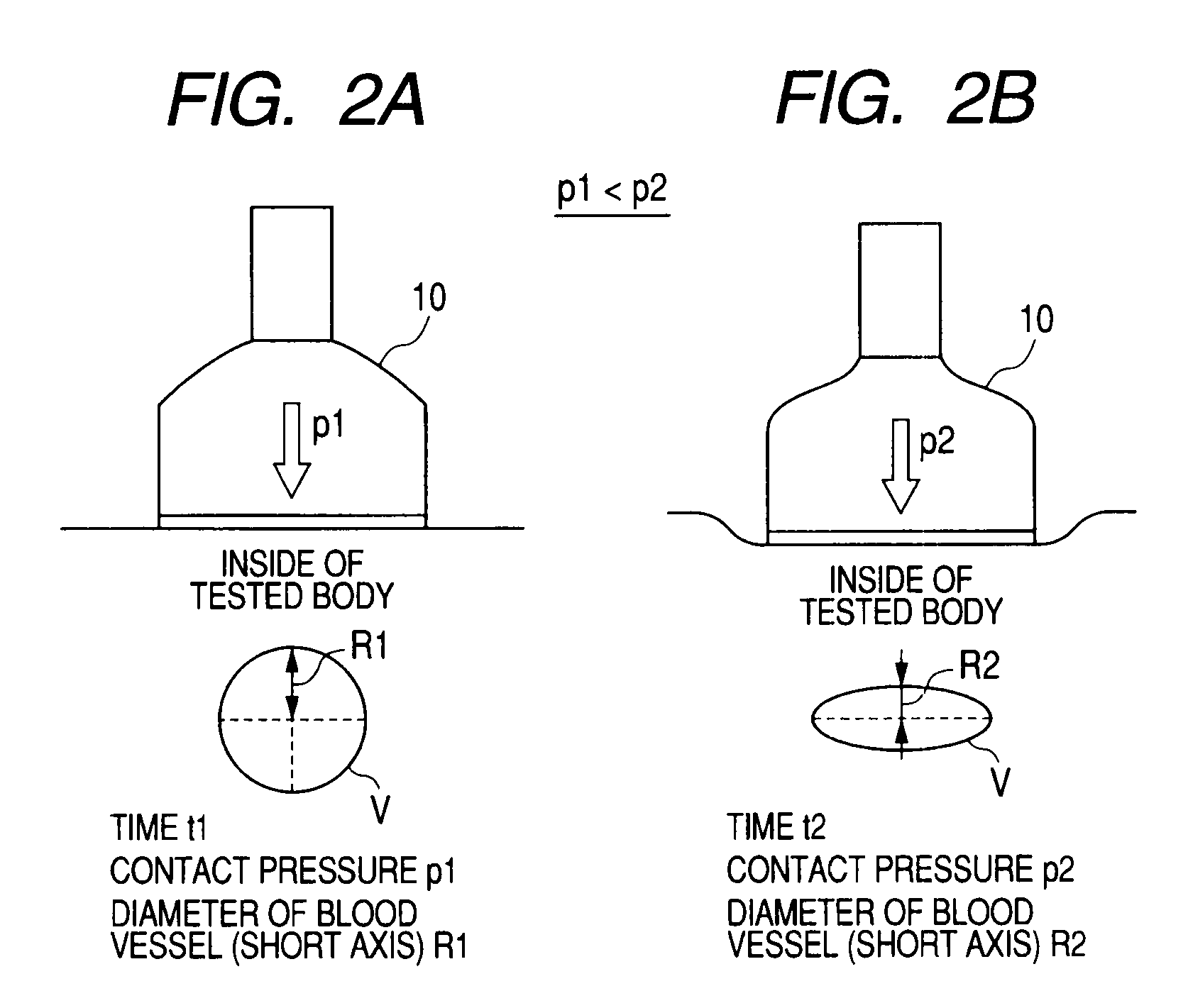
[0035]In a first embodiment, a pressure index value refers to a contact pressure between a probe and a tested body, which is caused by pressing the tested body with the probe.
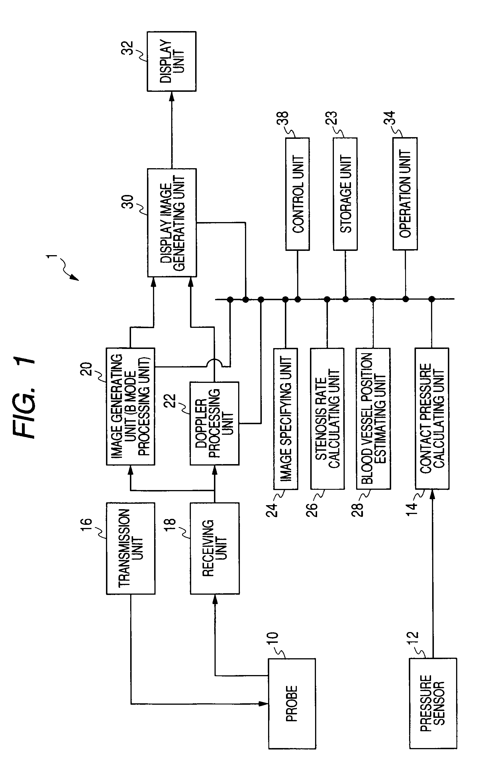
[0036]FIG. 1 is a view illustrating the configuration of an ultrasonic diagnostic apparatus 1 according to the first embodiment. As shown in the FIG. 1, the ultrasonic diagnostic apparatus 1 includes a probe 10, a pressure sensor 12, a contact pressure calculating unit 14, a transmission unit 16, a receiving unit 18, an image generating unit (B-mode processing unit) 20, a Doppler processing unit 22, a storage unit 23, an image specifying unit 24, a stenosis rate calculating unit 26, a blood vessel position estimating unit 28, a display image generating unit 30, a display unit 32, an operation unit 34, and a control unit 38. Hereinafter, each of the constituent components will be described.
[0037]The probe 10 has a vibrator array in which a plurality of vibrators for converting electric signals into ultrasonic wa...
second embodiment
[0106]In a second embodiment, a pressure index value is a Doppler shift frequency changing according to the movement of a blood vessel or a change of a blood flow that is caused by pressing a tested body with the probe 10. Moreover, in the following description, a component having approximately the same function as in the first embodiment is denoted by the same reference numeral, and repeat of explanation will only be made as needed.
[0107]FIG. 11 is a view illustrating the configuration of an ultrasonic diagnostic apparatus 50 according to a second embodiment of the invention. The ultrasonic diagnostic apparatus 50 includes a probe 10, a transmission unit 16, a receiving unit 18, an image generating unit (B-mode processing unit) 20, a Doppler shift frequency calculating unit (Doppler processing unit) 52, a storage unit 53, an image specifying unit 54, a stenosis rate calculating unit 26, a blood vessel position estimating unit 28, a display image generating unit 30, a display unit 3...
modified examples
[0142]In the first and second embodiments, examples using an ultrasonic image (B-mode image) on a two-dimensional tomographic plane have been explained. However, the invention does not need to be limited thereto but may also be applied to an ultrasonic image generated from volume data that is three-dimensional data. The ultrasonic image generated from volume data is assumed to be called a 3D image. In description of the following modified examples, a pressure index value is assumed to be a contact pressure in the same manner as in the first embodiment. However, in the modified examples, the pressure index value may also be a Doppler shift frequency.
[0143]The ultrasonic diagnostic apparatus 1 in the modified example repeatedly scans a three-dimensional region of a tested body with an ultrasonic wave by using a probe 10′.
[0144]An image generating unit 20′ reconstructs volume data on the basis of brightness data from the probe 10′ and generates data of the 3D image related to a predete...
PUM
 Abstract
Description
Claims
Application Information
 - R&D
- Intellectual Property
- Life Sciences
- Materials
- Tech Scout
- Unparalleled Data Quality
- Higher Quality Content
- 60% Fewer Hallucinations
Browse by: Latest US Patents, China's latest patents, Technical Efficacy Thesaurus, Application Domain, Technology Topic, Popular Technical Reports.
© 2025 PatSnap. All rights reserved.Legal|Privacy policy|Modern Slavery Act Transparency Statement|Sitemap|About US| Contact US: help@patsnap.com


