System and method for noninvasive electrocardiographic imaging (ECGI)

a non-invasive electrocardiographic and imaging technology, applied in the field of improvement, can solve the problems of reducing the applicability of bem ecgi to clinical applications, requiring large memory resources, and requiring iterative time-consuming tasks, so as to improve the turnaround time of ecgi, reduce complexity, and improve the effect of accuracy

Active Publication Date: 2011-07-19
CASE WESTERN RESERVE UNIV +1
View PDF10 Cites 154 Cited by
  • Summary
  • Abstract
  • Description
  • Claims
  • Application Information

AI Technical Summary

Benefits of technology

[0008]Rather than employing a surface mesh of the body surface and heart surface, MFS ECGI operates to define a plurality of virtual source nodes both outside the body surface and inside the heart surface. The virtual source nodes that are located outside the body surface define a surface boundary outside the body surface. The virtual source nodes that are located inside the heart surface define a surface boundary inside the heart surface. Based on the known geometrical relationships between the virtual source nodes, the electrode positions where the body surface potentials are measured, and the epicardial nodes for which the heart surface electrical potentials are computed, the MFS technique can readily reconstruct the epicardial cardiac surface potentials from the measured body surface potentials.
[0010]The inventors herein believe that the improved performance of MFS ECGI relative to BEM ECGI opens wide new windows of opportunity for noninvasive ECGI, particularly in connection with medical applications where time is of the essence such as interventional medical applications (including but not limited to ablation of arrhythmia substrates, targeted drug delivery, lead placement for implanted devices such as pacemakers and implanted cardioverters / defibrillators (ICDs), and other surgical procedures), guidance of interventional medical applications, evaluation of drug effect, risk stratification, and exercise stress tests. Even in less time critical applications, the inventors herein believe that the present invention will dramatically improve turnaround time for ECGI such that results can be obtained in minutes rather than hours, even while the patient remains in the cardiac electrophysiology laboratory, thereby allowing for rapid diagnosis and possible ECGI-guided intervention.
[0011]Moreover, because of the reduced complexity and increased automation of MFS ECGI relative to BEM ECGI, it is believed by the inventors herein that the amount of training required by a user (such as a physician, fellow, or medical assistant) can be greatly reduced, thereby allowing for wider use in the field

Problems solved by technology

This 3D surface meshing is an iterative time-consuming task that requires large memory resources.
However, this initial mesh will often need to be optimized to improve its accuracy, thereby further adding to the time required to accurately reconstruct the surface potentials and, in turn, further detracting from BEM ECGI's applicability to clinical applications.
Moreover, the skill level required to optimize body surface and heart surface meshes is generally high, which limits the pool of people who are qualified to conduct BEM ECGI.
Further still, even with a skilled person performing mesh optimization, it is believed by the inventors herein that BEM meshes nevertheless exhibit difficulty in accommodating complex heart geometries (particularly concave geometries) such as those that may be found in patients suffering from heart disease.

Method used


Image

  • System and method for noninvasive electrocardiographic imaging (ECGI)
  • System and method for noninvasive electrocardiographic imaging (ECGI)
  • System and method for noninvasive electrocardiographic imaging (ECGI)

Examples


Embodiment Construction

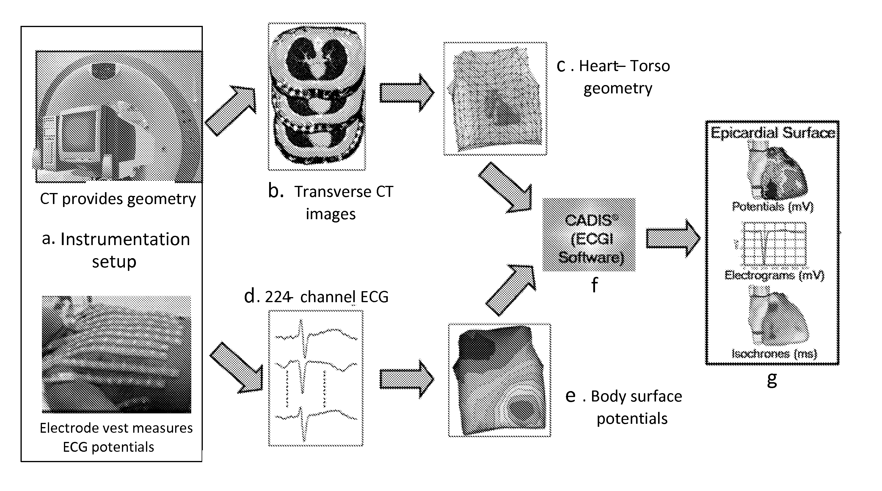
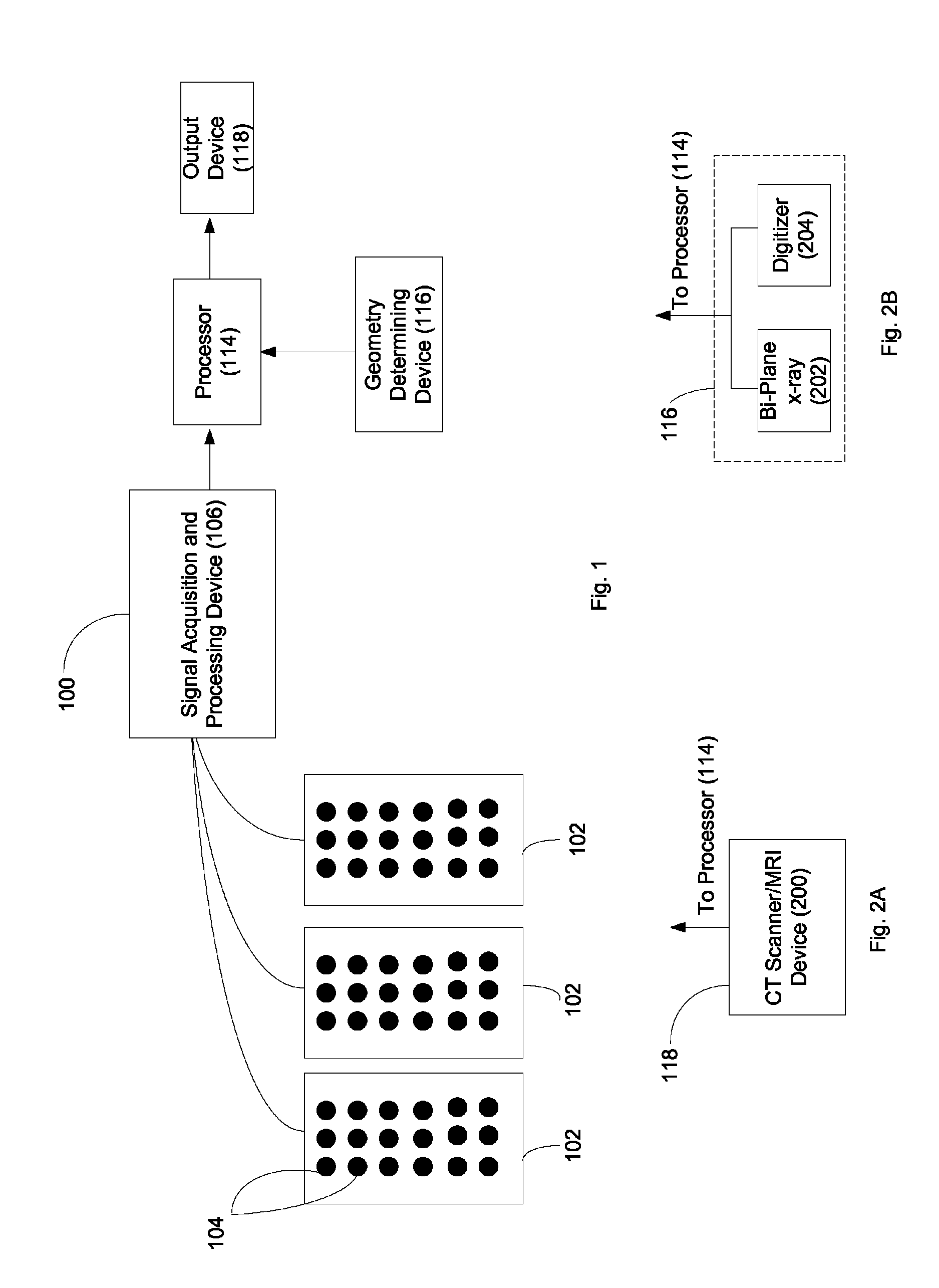
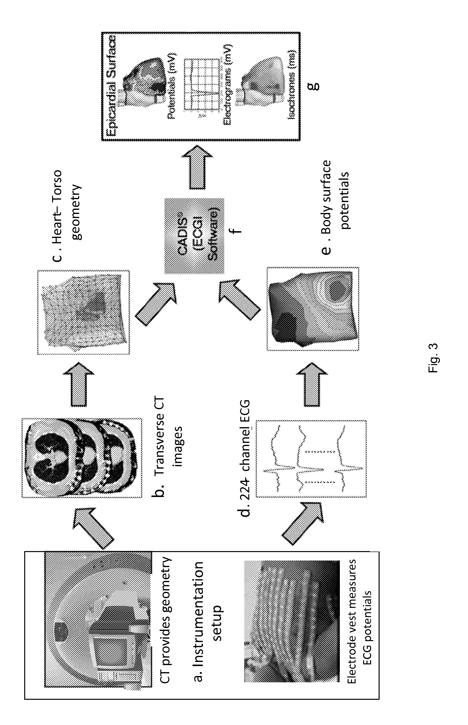
[0028]FIG. 1 depicts a block diagram overview of a preferred system 100 for performing meshless noninvasive ECGI. The system 100 preferably comprises a plurality of electrodes 104 (mounted on strips 102, a vest, or in some other array) in communication with a signal acquisition and processing device 106. The electrodes 104 serve to sense a plurality of electrical potentials on a patient's body surface. The signal acquisition and processing device 106 operates to process this sensed data to a form suitable for digital processing, as is known in the art. The system 100 also comprises a geometry determining device 116 that serves to generate data that is indicative of the geometrical relationship between the electrodes 104 and one or more points of interest within the patient (e.g., the patient's epicardial cardiac surface).

[0029]Processor 114 operates to (1) receive data from both the electrodes 104 (by way of the signal acquisition and processing device 106) and the geometry determin...


PUM


Abstract

Noninvasive systems and methods are provided for determining electrical activity for a heart of a living being. A processor is configured to meshlessly compute data that represents heart electrical activity from a set of noninvasively measured body surface electrical potentials. This is accomplished using data that describes a geometric relationship between a plurality of locations corresponding to where the body surface electrical potentials were measured and the heart.

Description

RELATED APPLICATION[0001]The present application claims priority to U.S. Provisional Application No. 60 / 701,626 file Jul. 22, 2005, which is incorporated herein by reference in its entirety.STATEMENT REGARDING FEDERALLY SPONSORED RESEARCH OR DEVELOPMENT[0002]This invention was made with government support under NIH-NHLBI Grant R37-HL-33343 awarded by the National Institutes of Health (NIH). The government may have certain rights in the inventionFIELD OF THE INVENTION[0003]This invention relates to an improved technique for noninvasive electrocardiographic imaging (ECGI). In particular, the preferred embodiment of the present invention relates to a meshless noninvasive ECGI technique wherein a plurality of body surface potentials are noninvasively obtained and combined with data representing the geometry of a heart and body torso to generate electrocardiographic images that represent electrical activity of the heart.BACKGROUND OF THE INVENTION[0004]Over 7 million people worldwide (ar...

Claims


Application Information

Patent Timeline
Patent Type & Authority Patents(United States)
IPC IPC(8): A61B5/04
CPCA61L2/10A61L2/202A61L9/015A61L9/20A61B5/055A61B6/03A61B5/0044A61B5/282A61B5/318
Inventor RUDY, YORAMWANG, YONGJIA, PING
Owner CASE WESTERN RESERVE UNIV
Why Eureka
Try Eureka