
Vertebral disc repair

a technology of vertebral discs and implants, applied in the field of shaped implants, can solve the problems of debilitating lower back and neck pain, np cannot no longer effectively transfer loads, af layers do not have the same ability to resist compressive loads, and experience atypical stresses, etc., to achieve the effect of reducing osteoinductivity

Inactive Publication Date: 2011-02-01
MUSCULOSKELETAL TRANSPLANT FOUND INC
View PDF226 Cites 96 Cited by
  • Summary
  • Abstract
  • Description
  • Claims
  • Application Information

AI Technical Summary

Benefits of technology

The invention is about a new type of implant for repairing a damaged spinal disc. The implant is made from a type of bone that has been treated to remove its ability to grow new bone cells. This makes it safer and more effective for use in the body. The implant can be shaped into a variety of forms and is designed to fit between the vertebrae in the spine and act as a cushion to reduce pressure and protect the spinal column. The invention can be made from either cortical or cancellous bone and can be sterilized and compressed for implantation. Overall, the invention provides a better option for repairing damaged spinal discs and promoting spinal health.

Problems solved by technology

Degeneration of the intervertebral disc within the spine is generally believed to be a common cause of debilitating lower back and neck pain.
With these pathologic changes, the NP can no longer effectively transfer loads and provide sufficient pressurization to keep the AF in tension.
When not properly tensioned, the layers of the AF do not have the same ability to resist compressive loads and experience atypical stresses.
Without a healthy NP to resist the AF from bulging inward, this abnormal stretching of the AF causes this tissue structure to weaken by making the successive plies buckle and separate from each other.
This causes the AF to become more susceptible to radial fissures or cracks under loading.
Over time, the disc also loses stability and height bringing the spinal facet joints in close contact with each other.
This phenomenon is often referred to as “herniated” disc with the nerve impingement typically resulting in debilitating back or leg pain, loss of muscle control or even paralysis.
The most common resulting symptoms are pain radiating along a compressed nerve and low back pain, either of which can be crippling for the patient.
When conservative approaches are not successful, the most common surgical options are currently discectomy and spinal fusion.
While both of these options are reasonably successful at acutely decreasing pain, neither one restores proper biomechanics to the spine, which may lead to further degeneration at the operated disc or discs at the adjacent levels in the spine.
The most significant drawbacks of discectomy are recurrence of herniation, recurrence of radicular symptoms, continuing loss of disc height and increasing low back pain.
All of these failings are most directly related to the loss of NP material and AF competence that results from herniation and surgery.
Loss of NP material via discectomy further deflates the disc, causing a decrease in disc height.
Loss of disc height increases loading on the facet joints.
This can result in deterioration of facet cartilage and ultimately osteoarthritis and pain in this joint.
This leads to foraminal stenosis, pinching of the traversing nerve root, and recurring radicular pain.
Loss of NP also increases loading on the remaining AF, a partially ennervated structure that can produce pain.
Finally, loss of NP results in greater bulging of the AF under load.
This can result in renewed impingement by the AF on nerve structures posterior to the disc.
Persisting tears in the AF that result either from herniation or surgical incision also contribute to poor results from discectomy.
This may over-stress the facets and contribute to their deterioration.
In these cases, not only is re-herniation a risk but also leakage of fluids or solids from within the NP into the epidural space can occur.
This has been shown to cause localized pain, irritation of spinal nerve roots, decreases in nerve conduction velocity, and may contribute to the formation of post-surgical scar tissue in the epidural space.
If successful, this results in the two vertebrae being “fused” together This treatment generally reduces back pain but limits the mobility of the spine.
It is suspected that this abnormal biomechanical loading may lead to DDD and repeat surgeries at the adjacent levels.
All present surgical interventions, whether laminectomy or fusion of adjacent vertebrae, lower the functionality of the spine in some way.
This is, however, extremely difficult.
Beyond the questionable efficacy of those devices were the inherent difficulties encountered during implantation.
These mechanical discs are in the early stages of clinical evaluation and are relatively unproven.
These plastic debris particles have caused serious complications in the knee and hip applications.
The construction of a fully functional prosthesis is extremely difficult and most prosthetic devices suggested to date are strictly mechanical, and they mimic only some functions of the disc.
One disadvantage of this solution, which is common to all full spinal disc replacements, remains a complicated surgical procedure, which translates into a high cost, and a high risk to the patient.
These solid implants did not restore proper biomechanics in discs due to their stiffness.
Unfortunately, however, most prosthetic nucleus devices that are designed to be implanted through a small annulotomy do not properly fill the nuclear cavity.
Degenerated, painfully disabling spinal discs are a major economic and social problem for patients, their families, employers and the public at large.
Other means to replace the function of a degenerated disc have major problems such as complex surgical procedures, unproven efficacy, placing unnecessary and possibly destructive forces on an already damaged annulus, etc.

Method used


Image

  • Vertebral disc repair
  • Vertebral disc repair
  • Vertebral disc repair

Examples


Embodiment Construction

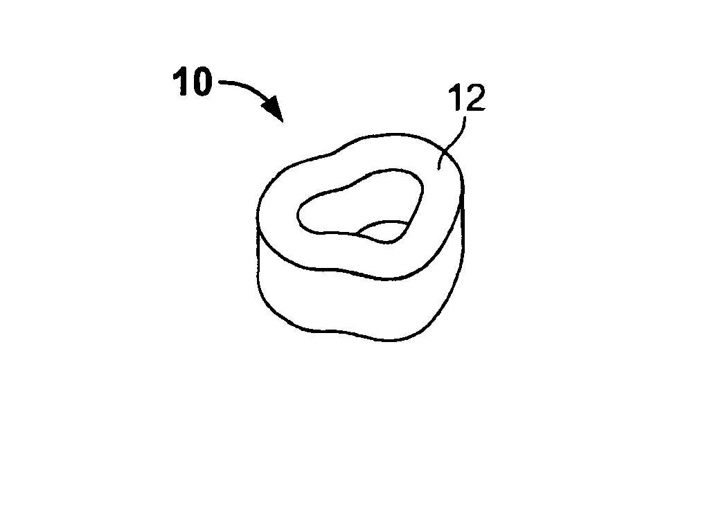
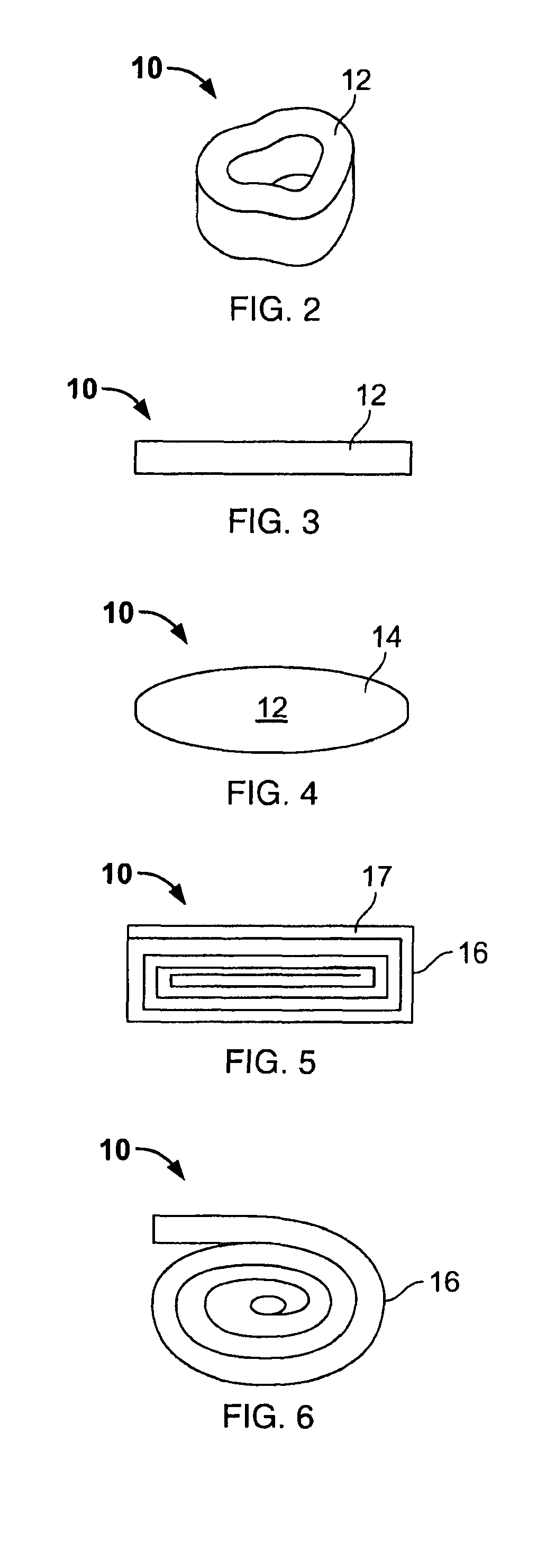
[0051]While the present invention is susceptible of embodiment in various forms as is shown in the drawings, and will hereinafter be described, a presently preferred embodiment is set forth with the understanding that the present disclosure is to be considered as an exemplification of the invention, and is not intended to limit the invention to the specific embodiments disclosed herein. The preferred embodiment and best mode of the invention for these purposes is shown in FIGS. 2 through 4.

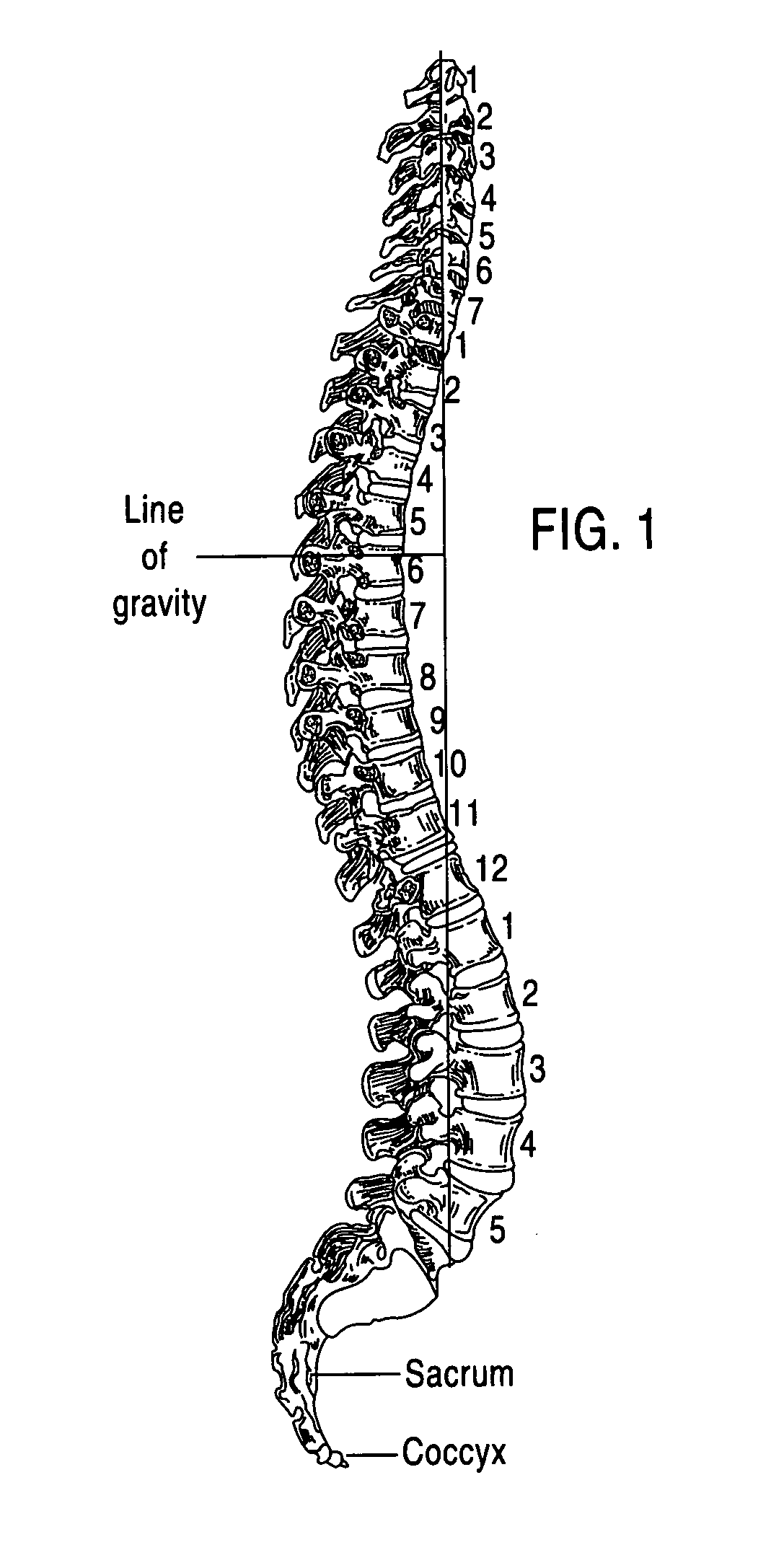
[0052]The present invention is directed toward a spinal disc repair implant fashioned from demineralized human allograft bone and more particularly toward an implant 10 that includes at least one load bearing elastic body 12 sized for introduction into an intervertebral disc space as shown in FIG. 1. FIG. 1 shows a spinal column with numbered vertebrae separated by discs. The implants have shape memory and are configured to have a specific original shape that allows extensive deformation without p...


PUM

PropertyMeasurementUnit
diameteraaaaaaaaaa
thickaaaaaaaaaa
heightaaaaaaaaaa

Abstract

A sterile implant for treatment of a spinal disc defect comprising an allograft cortical bone demineralized to a Type I collagen having a specific shape which is treated to eliminate osteoinductivity. The implant is lyophilized and compressed into smaller first shape which 20 to 80% from its original shape in at least one dimension and hardened. The implant expanding when hydrated into a second shape having the shape memory of the first shape and expanded in dimensional size from the first compressed shape.

Description

RELATED APPLICATIONS[0001]This is an application claiming priority from U.S. Provisional Application No. 60 / 671,514 filed Apr. 15, 2005 and U.S. Provisional Application No. 60 / 791,904, filed Apr. 14, 2006.FIELD OF INVENTION[0002]The present invention is directed toward a shaped implant constructed of Type I collagen obtained from demineralized bone which is used for human spinal disc repair. The Type I collagen is treated to eliminate osteoinductivity and the implant is used to replace or augment the nucleus pulposus of a degenerated spinal disc after rupture or herniation. More specifically, the present invention is directed to a load-bearing implant which possesses a unique advantage of shape-memory.BACKGROUND OF THE INVENTION[0003]Degeneration of the intervertebral disc within the spine is generally believed to be a common cause of debilitating lower back and neck pain. An intervertebral disc primarily serves as a mechanical cushion between the vertebral bones, permitting control...

Claims


Application Information

Patent Timeline
Patent Type & Authority Patents(United States)
IPC IPC(8): A61F2/44
CPCA61F2/28A61F2/442A61L27/3608A61L27/3658A61L27/38A61B17/842A61F2002/30057A61F2002/30059A61F2002/30062A61F2002/30075A61F2002/3008A61F2002/30136A61F2002/30153A61F2002/30172A61F2002/30177A61F2002/30181A61F2002/302A61F2002/30224A61F2002/30225A61F2002/30242A61F2002/30293A61F2002/30383A61F2002/30387A61F2002/30795A61F2002/30932A61F2002/30971A61F2002/4435A61F2002/444A61F2210/0004A61F2210/0061A61F2220/0025A61F2230/0004A61F2230/0019A61F2230/0052A61F2230/0056A61F2230/006A61F2230/0065A61F2230/0069A61F2230/0071A61F2230/0091A61F2250/0098A61F2310/00365A61L2400/16A61L2430/38Y10S623/919
Inventor GERTZMAN, ARTHUR A.MERBOTH, BARBARA L.SCHULER, MICHAELSTEINER, ANTON J.SEMLER, ERIC J.YANNARIELLO-BROWN, JUDITH I.
Owner MUSCULOSKELETAL TRANSPLANT FOUND INC
Learn More