Method for reducing exposure to infrared light beam, ultrasound or magnetic impulse rays in medical imaging devices

a technology of infrared light beam and magnetic impulse ray, which is applied in the direction of material analysis, material analysis using wave/particle radiation, instruments, etc., can solve the problem that the length of time the system is used for a specific patient cannot exceed a certain economic threshold, and achieve the effect of increasing the overall radiation dos

Inactive Publication Date: 2010-09-14
BEYRARD NORBERT
View PDF11 Cites 5 Cited by
  • Summary
  • Abstract
  • Description
  • Claims
  • Application Information

AI Technical Summary

Benefits of technology

[0046]The early detection of cancer presupposes a considerable gain in definition. However, the length of time the system is used for a specific patient cannot exceed a certain economic threshold. Above all however, increasing the number of profiles increases the overall radiation dose.
[0049]To this end, the object of the present invention is a method intended to reduce exposure to radiation or to ultrasonic beams or magnetic pulse beams in an organism, from medical imaging equipment, thanks to a modification of the procedure for processing signals, in order to obtain representative images of values associated with each point or zone of a planar or non-planar surface, which comprises:
[0050]producing a first image by known methods in order to obtain as precise an estimation as possible of the value associated with each point or zone in a representation of the image, via a rectangular matrix, by evaluating by interpolation values between two points or by distributing the value of a zone into micro-zones obtained by splitting up the zone, so as to arrive at an amplified matrix starting from the initial matrix, thereby giving a more accurate image of the initial image,
[0062]Each of the terms Bij is evaluated from a set of physical scannings spaced so as to produce a reduced matrix representative of the image, by distributing each of the terms of the reduced matrix in a square in which the number of lines and columns is equal to k, regarded as the coefficient of amplification of the initial matrix, so as to obtain the reference matrix, amplified k times, and resulting in a more accurate first image, intended to be adjusted.

Problems solved by technology

However, the length of time the system is used for a specific patient cannot exceed a certain economic threshold.

Method used


Image

  • Method for reducing exposure to infrared light beam, ultrasound or magnetic impulse rays in medical imaging devices
  • Method for reducing exposure to infrared light beam, ultrasound or magnetic impulse rays in medical imaging devices
  • Method for reducing exposure to infrared light beam, ultrasound or magnetic impulse rays in medical imaging devices

Examples


Embodiment Construction

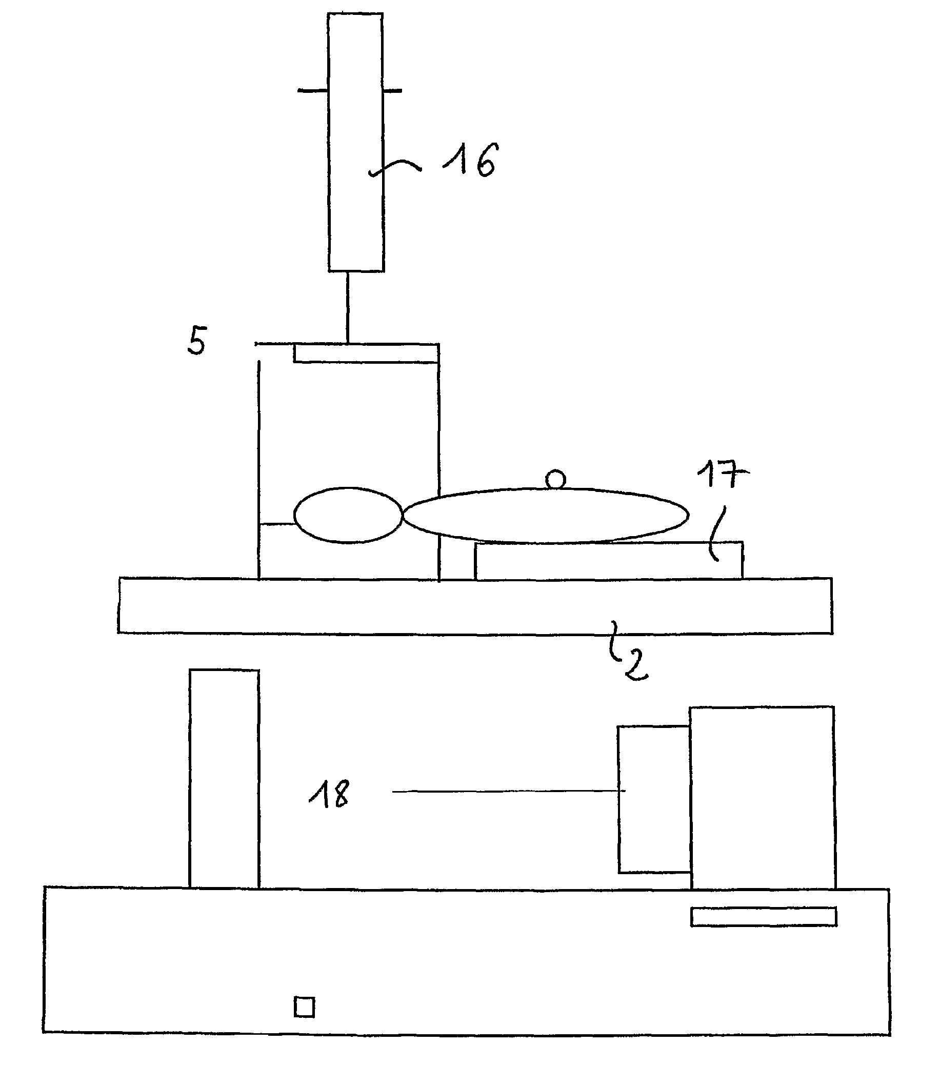
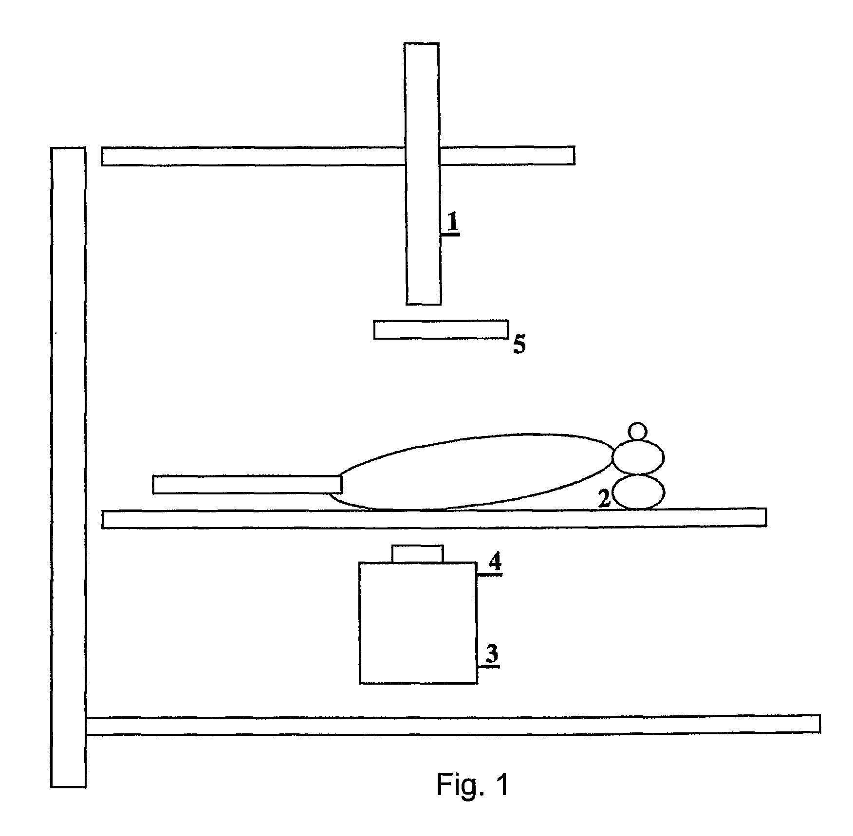
[0090]In a preferred embodiment in which a laser or infra-red beam is used, two transparent plates are provided, coated with anti-reflection layers in order to prevent a reflection of the infra-red beam, and mutually parallel in order to limit a refraction of this beam, the object to be examined being arranged between the two plates and resting against one of them serving as support plates.

[0091]Thus, the combination of an accurate scanning apparatus together with screens of very small dimensions that can be positioned and switched in an extremely precise manner, and an information processing system providing an acceptable evaluation of the values of the coefficient of attenuation of each elementary micro-zone, enables the number of beam pulses and thus the number of profiles to be recorded to be reduced by a considerable factor, while at the same time increasing the definition from 1 mm to 10 or 20 microns without involving a prohibitive level of irradiation.

[0092]In fact, for a 10...


PUM


Abstract

Method and apparatus for X-ray imaging of a body, employing a support to receive a body to be examined, a source emitting a beam of X-rays, a detector irradiated by the beam, a converter for converting the detected intensities into data, a means for turning the mounted mobile support by an angle of rotation about an axis of rotation with respect to the source and the detector and a suitably programmed computer to average the data acquired for a pair of orthogonal angles of rotation to obtain n column and m line mean values for n and m elementary segments of a band of the detector, to construct an initial image (n, m) with the n column and m line mean values, to adjust the coefficient of attenuation in each n×m elementary zone by a method of least squares taking into account the n column and m line mean values regarded as constraints, to repeat the previous stages for data acquired with different pairs of preferably orthogonal angles of rotation, and to average term by term the adjusted images so as to arrive at a synthesis image expressing coefficients of attenuation of the examined body.

Description

CROSS REFERENCE TO RELATED APPLICATIONS[0001]This application is a Continuation application of PCT / FR2005 / 001437 filed Jun. 10, 2005, which claims priority of Application Nos. FR0406497 filed Jun. 16, 2004; FR0452677 filed Nov. 18, 2004, which are all incorporated by reference herein in their entirety.BACKGROUND OF THE INVENTION[0002]1. Field of the Invention[0003]The present invention relates to a method intended to reduce the exposure of an organism to radiation and infra-red, ultrasonic or magnetic pulse beams from medical imaging equipment, and comprises a method for treating a signal or a set of signals with a view to producing under greatly improved conditions digital images of the interior of an organism, in order to detect and treat where appropriate abnormalities or disorders in the examined organism.[0004]2. Description of the Related Art[0005]Various methods are now commonly used for this purpose, in particular radiography, scanography, echography and magnetic resonance i...

Claims


Application Information

Patent Timeline
Patent Type & Authority Patents(United States)
IPC IPC(8): G06K9/00
CPCA61B5/0091A61B5/4312A61B6/032A61B8/14
Inventor BEYRARD, NORBERT
Owner BEYRARD NORBERT
Why Eureka
Try Eureka