
Detection and monitoring of changes in mineralized tissues or calcified deposits by optical coherence tomography and Raman spectroscopy

a technology of which is applied in the field of detection and monitoring of changes in mineralized tissues or calcified deposits by optical coherence tomography and raman spectroscopy, can solve the problems of affecting the prevalence of dental caries in western countries, and affecting the normal function of these tissues. , to achieve the effect of improving sensitivity and specificity

Inactive Publication Date: 2010-09-14
NAT RES COUNCIL OF CANADA +2
View PDF6 Cites 126 Cited by
  • Summary
  • Abstract
  • Description
  • Claims
  • Application Information

AI Technical Summary

Benefits of technology

[0056]Preferably the light energy is delivered to the sample and signal collected from the sample via at least one fibre optic cables interfaced with an ergonomically-shaped dental handpiece probe with an intra-oral portion and wherein the probe contains a probe head and tip arranged to enable imaging / measurements in the gingival embrasure of the interproximal spaces between adjacent teeth, the tip having a triangular shaped tip with a triangular cross-section to allow the tip to be wedged into tight spaces such as the gingival embrasures between teeth, the tip being formed of a material that allows transmission and collection of light energy with minimal disruption.
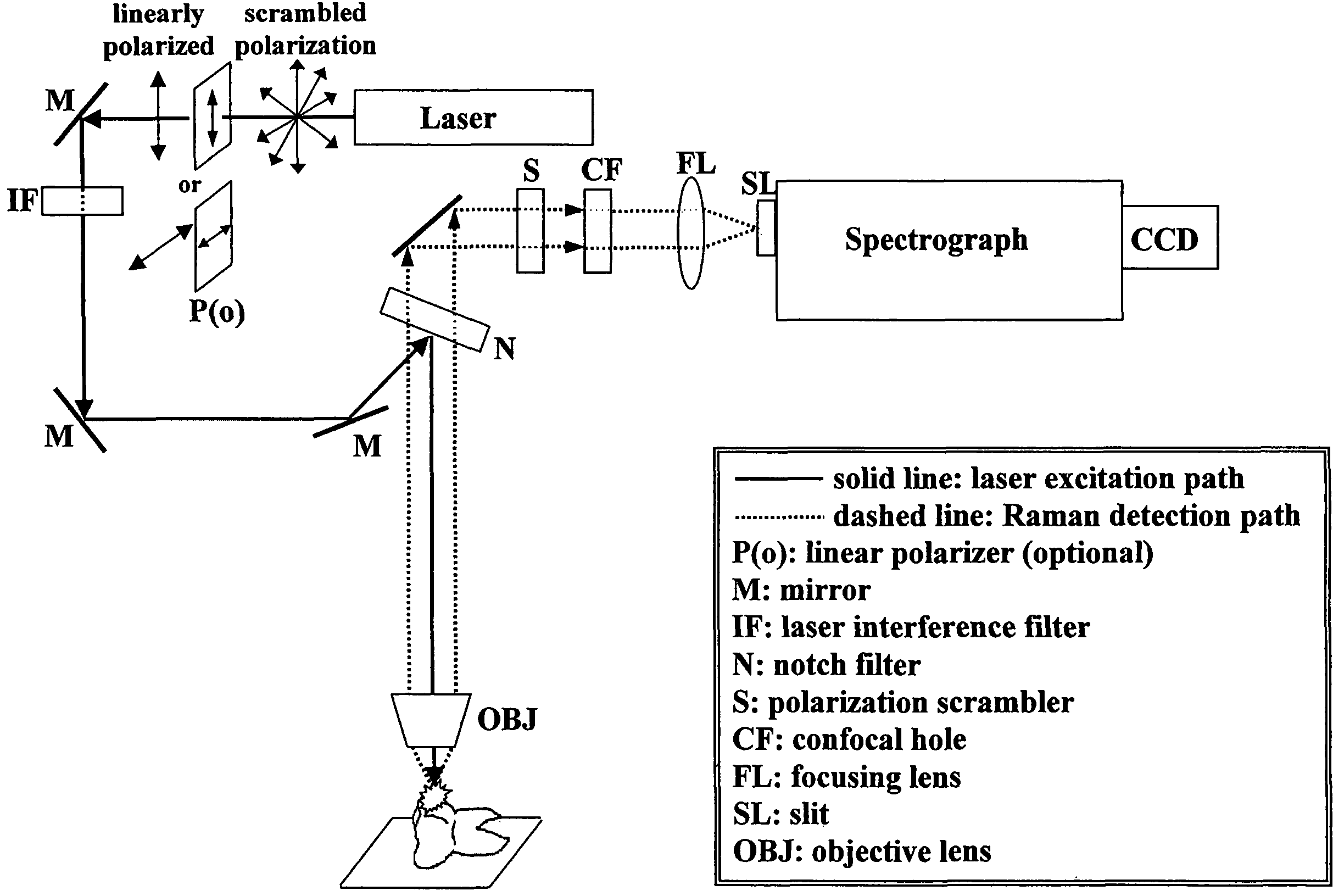
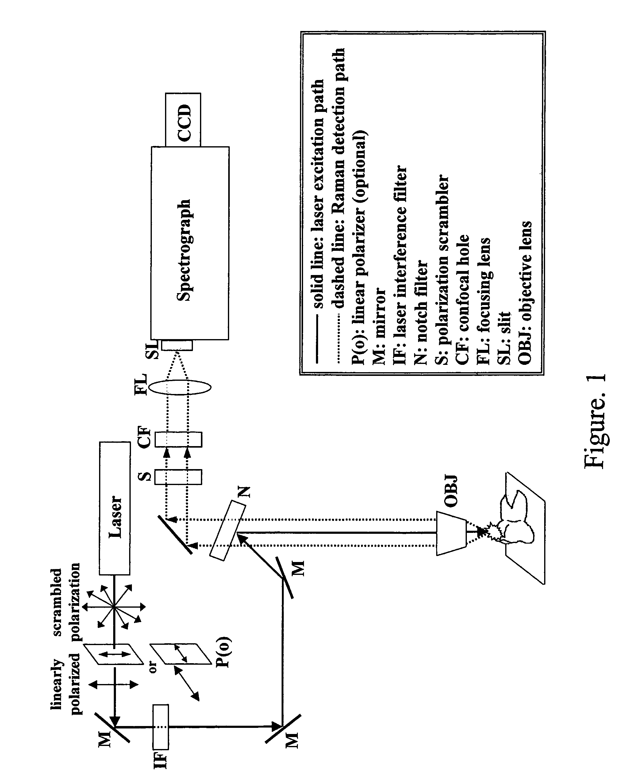
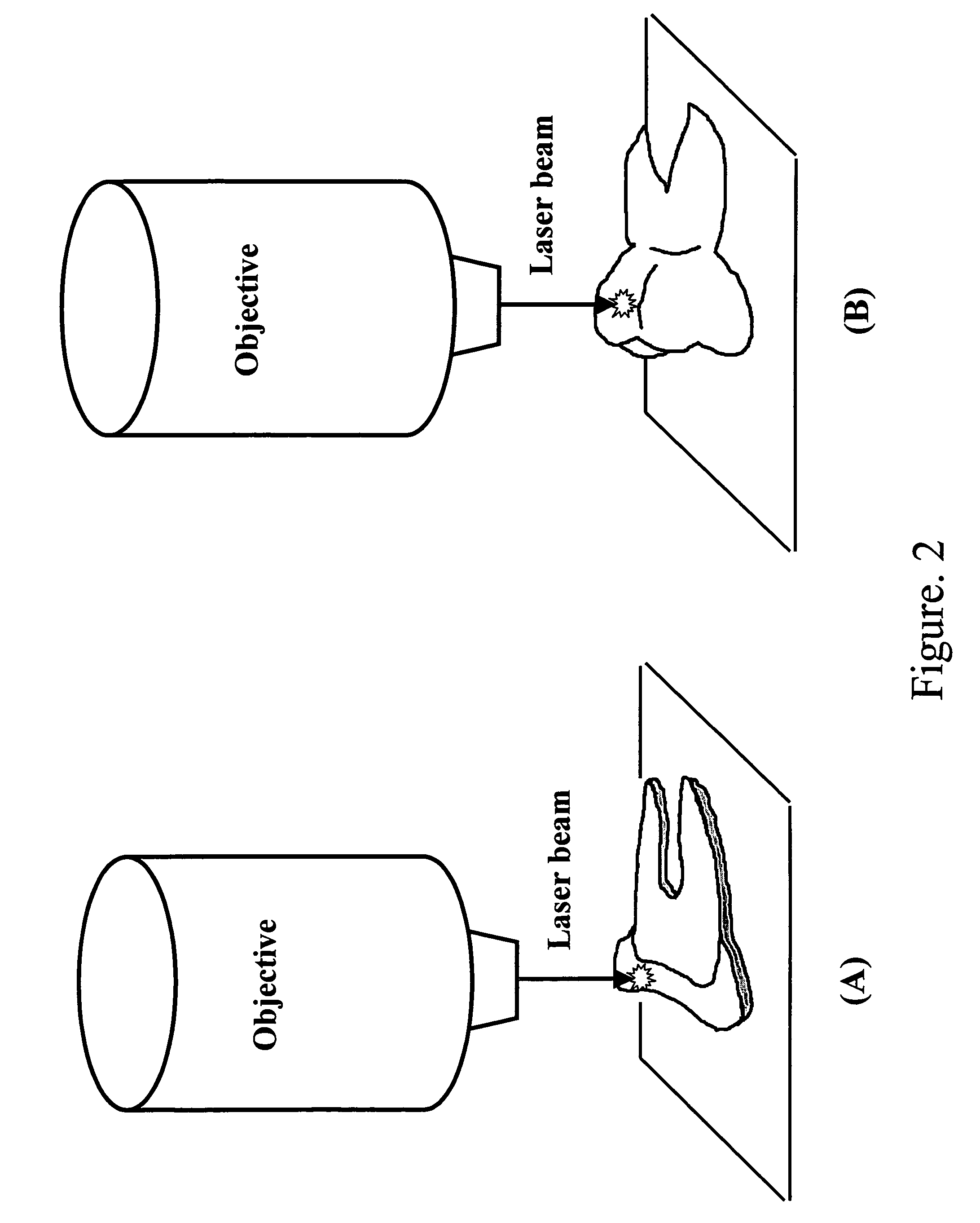
[0101]Various studies using OCT and Raman spectroscopy are provided to detect and characterize early enamel carious lesions in extracted human teeth. Comparisons will be made with results obtained from sound tooth enamel. The Raman studies are conducted with microspectroscopy, polarized Raman spectroscopy and fibre-optic sampling to illustrate the similarity of the information obtained with both (microscopy and fibre-optic) measurement modes and the potential of developing the Raman technique combined with OCT, into a fibre based tool for clinical applications with improved sensitivity and specificity.

Problems solved by technology

Such deposits can disrupt the normal function of these tissues.
However, in diseased states such as upon the attack of acid by acid-forming bacteria, the ordered structure is disturbed creating porous regions and amorphous areas.
With the introduction of fluoride in drinking water and toothpastes, there has been a decline in the prevalence of dental caries in Western countries.1 However, a recent report states that caries still remains a large problem in specific population groups (e.g. minority children, the economically underprivileged, older people, the chronically ill and institutionalized persons).2 Furthermore, the patterns of caries development are changing, to those with smaller lesion sizes and slower progression rates, making caries more difficult to detect with existing conventional techniques.1 This is further complicated when caries develop at locations which are not clinically visible (e.g. between adjacent teeth).
Such methods may not be reliable for detecting interproximal lesions (those between adjacent teeth) because these carious lesions are not clinically visible.
In addition, these clinical methods do not adequately detect changes in caries development and do not possess the sensitivity, specificity nor ability to account for the dynamic process of demineralization-remineralization.1,2 Therefore, more refined diagnostic tools are required to identify early non-cavitated carious lesions and to monitor their activity as well as severity.
Although the technique uses a lower radiation dose, the application of the method is still limited by the ionizing radiation required to obtain an image.
The drawback of the technique is its lack of specificity, which limits its usefulness in deciding whether or not to treat a lesion operatively.
With this method, the presence of bacterial plaque or saliva interferes with the usefulness of the technique.8 A related approach, laser fluorescence, uses light at 633 nm and the reflected fluorescence is measured.
Recent clinical studies indicate that this latter technique can lead to false-positive results due to the presence of stains, deposits, calculus and organic material in the region of interest9 and is not suitable for detecting initial carious changes in enamel.10 Therefore, despite the potential of these various technologies, the methods are prone to subjectivity issues with high intra- and inter-examiner variability, false-positive results due to stains or organic deposits and the unsuitability for detecting initial enamel caries at interproximal sites.

Method used


Image

  • Detection and monitoring of changes in mineralized tissues or calcified deposits by optical coherence tomography and Raman spectroscopy
  • Detection and monitoring of changes in mineralized tissues or calcified deposits by optical coherence tomography and Raman spectroscopy
  • Detection and monitoring of changes in mineralized tissues or calcified deposits by optical coherence tomography and Raman spectroscopy

Examples


Embodiment Construction

[0133]Human molars and premolars (n=15) were collected from orthodontic patients at the Dental Clinic of the Faculty of Dentistry, University of Manitoba. Approvals from the human ethics committees of the Institute for Biodiagnostics (National Research Council Canada), University of Manitoba and Dalhousie University were obtained prior to sample collection. All teeth were examined in patients before extraction and no discoloration of the marginal ridge was observed. Remaining soft tissue on extracted teeth was removed by scaling and the samples were thoroughly rinsed with water. Teeth were preserved in sterile filtered de-ionized water until measurement. Each tooth sample was radiographed and independently re-assessed ex vivo by two clinical investigators at the University of Manitoba and Dalhousie University, respectively. Control caries-free teeth had no visible decalcification or demineralization. Incipient carious teeth included regions of decalcification with intact surfaces an...


PUM

PropertyMeasurementUnit
wavelengthaaaaaaaaaa
excitation wavelengthsaaaaaaaaaa
porosityaaaaaaaaaa

Abstract

Early dental caries detection is carried out by a method that combines optical coherence tomography (OCT) and Raman spectroscopy to provide morphological information and biochemical specificity for detecting and characterizing incipient carious lesions found in extracted human teeth. OCT imaging of tooth samples demonstrated increased light back-scattering intensity at sites of carious lesions as compared to the sound enamel. Raman microspectroscopy and fibre-optic based Raman spectroscopy are used to characterize the caries further by detecting demineralization-induced alterations of enamel crystallite morphology and / or orientation. OCT imaging is useful for screening carious sites and determining lesion depth, with Raman spectroscopy providing biochemical confirmation of caries. The combination is incorporated into a common probe operable without movement to scan the tooth surface and to provide an output for the dentist.

Description

[0001]This application claims priority under 35 U.S.C. 119 from U.S. Provisional Application Ser. No. 60 / 578,070 filed Jun. 9, 2004.[0002]This invention relates to a method for detecting, monitoring and assessing changes in mineralized tissues or calcified deposits by optical coherence tomography and Raman spectroscopy. The invention is primarily but not exclusively designed for use in detecting early caries in tooth structures.BACKGROUND OF THE INVENTION[0003]Calcification is the process whereby the mineral calcium builds up in tissue, causing it to harden. Calcium that enters the body is normally deposited in bones and teeth (i.e. mineralized tissues). Ossified (hardened) tissues such as bone and teeth are composed of calcium phosphate and calcium carbonate deposits. In particular, the bulk of the mineral phase of bones and teeth is a poorly crystalline carbonate-rich analogue of the naturally occurring mineral hydroxyapaptite. When there is an imbalance in the body, calcium and o...

Claims


Application Information

Patent Timeline
Patent Type & Authority Patents(United States)
IPC IPC(8): G01N21/00A61B5/00
CPCA61B5/0066A61B5/0088
Inventor CHOO-SMITH, LIN-P'INGKO, ALEX C. T.HEWKO, MARKLEONARDI, LORENZOCLEGHORN, BLAINEDONG, CECILIA
Owner NAT RES COUNCIL OF CANADA
Learn More