
Method and apparatus for analyzing biological tissue specimens

Inactive Publication Date: 2009-09-29
HUMANITAS MIRASOLE S P S +1
View PDF4 Cites 4 Cited by
  • Summary
  • Abstract
  • Description
  • Claims
  • Application Information

AI Technical Summary

Problems solved by technology

In any case, several drawbacks are however present in the known apparatuses.
The main drawback concerns the way the acquired image is processed by the computer.
In such a case, the known devices do not allow a correct quantification of the requested parameters (perimeter, area, etc.) to be made, particularly for highly irregularly shaped objects such as the ones named above, with the consequence that the outcome of the analysis may be incorrect or even misleading.
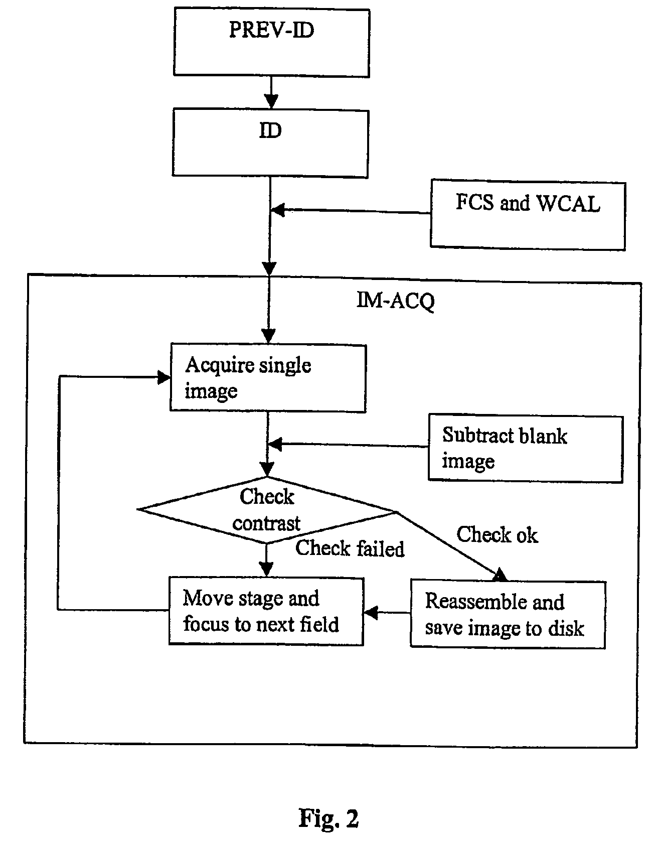
Another typical problem in the case of observation through microscope is the automatic focusing of the image.
In fact, when at high magnification a scanning of the slide is required in order to observe or capture the overall image to be analyzed, a fast and accurate focusing of each scanned part is difficult to be performed.

Method used


Image

  • Method and apparatus for analyzing biological tissue specimens
  • Method and apparatus for analyzing biological tissue specimens
  • Method and apparatus for analyzing biological tissue specimens

Examples


Embodiment Construction

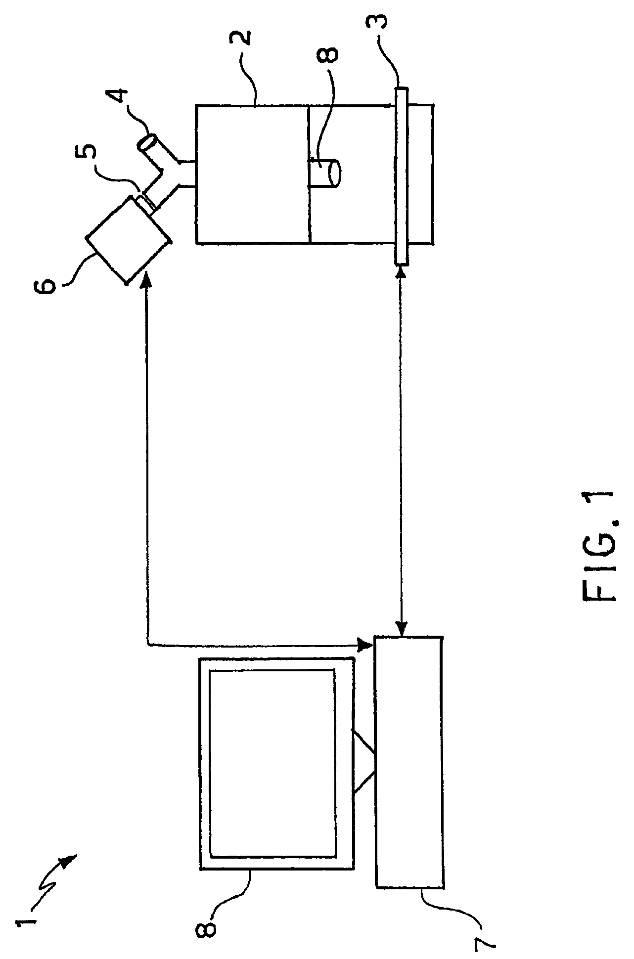
[0017]The example that will be described hereinafter concerns a system 1 for acquiring and processing an image comprising a microscope 2 having a motorized scanning stage 3 capable of moving along the Cartesian axis x, y, z. The microscope 2 is preferably of the type that allow magnification of from 50× up to 1000×.

[0018]The microscope 2 is provided with at least one object glass 8, at least one eyepiece 4 and at least one photo-video port 5 for camera attachment. To this latter, electronic image acquisition means 6, in particular a photo / video camera, are operatively connected. Preferably, such electronic image acquisition means 6 are a digital camera, having more preferably a resolution of 1.3 Megapixels.

[0019]The electronic image acquisition means 6 are operatively connected with a processing system 7. The processing system 7 may be realized by means of a personal computer (PC) comprising a bus which interconnects a processing means, for example a central processing unit (CPU), t...


PUM


Abstract

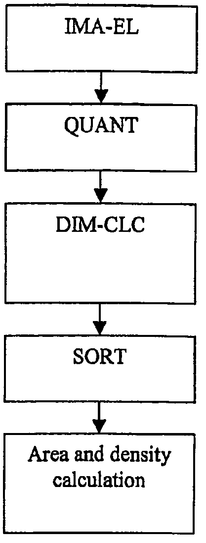
The present invention relates to a method and an apparatus for processing images of irregularly shaped objects, such as biological specimens, in particular of human or animal origin, or images thereof. The metric quantification of a biological body part or tissue or of an abnormal material spot or aggregate contained therein is also performed by means of the invention method. In particular, the present invention relates to a method for processing images of irregularly shaped objects, comprising a stage of acquisition of a digital image of said object, a stage of image elaboration (IMAEL) for quantifying said digital image to 1 bit and a stage of metrical processing of said 1-bit quantized image, wherein said stage of metrical processing comprises: —) a stage of object's metrical quantification (QUANT) for determining Euclidean perimeter P and / or area A of said object; —) a stage of dimensional calculation (DIM-CLC) for calculating a fractal-corrected perimeter Pf and / or area Af of said object.

Description

BACKGROUND OF THE INVENTION[0001]1. Field of the Invention[0002]The present invention relates to a method and an apparatus for processing images of irregularly shaped objects, such as biological specimens, in particular of human or animal origin, or images thereof. The metric quantification of a biological body part or tissue or of an abnormal material spot or aggregate contained therein is also performed by means of the invention method.[0003]With the term “abnormal material spot or aggregate” it is intended a material spot or aggregate morphologically connected with a pathological condition or a condition which gives rise to a pre- or post-pathological situation. Examples of abnormal material spot or aggregate may be tumors, atherosclerotic plaques, edemas, hematomes, acute or chronic inflammatory lesions, scars and collagen diseases.[0004]2. Description of Related Art[0005]Observation and analysis of human, animal or plant tissues is normally performed by means of a microscope. W...

Claims


Application Information

Patent Timeline
IPC IPC(8): G06K9/00G06K9/46G06T1/00G06T7/00G06T7/60G06V10/42
CPCG06K9/00127G06T7/602G06T7/0012G06T2207/30024G06T7/62G06V20/69G06V10/42
Inventor DIOGUARDI, NICOLAGRIZZI, FABIORUSSO, CARLOFRANCESCHINI, BARBARA
Owner HUMANITAS MIRASOLE S P S
Learn More