Methods and compositions for preserving the viability of photoreceptor cells
a technology of photoreceptor cells and compositions, applied in the direction of drug compositions, peptide sources, peptide/protein ingredients, etc., can solve problems such as irreversible visual dysfunction, and achieve the effect of maintaining the viability of photoreceptor cells and preserving vision
- Summary
- Abstract
- Description
- Claims
- Application Information
AI Technical Summary
Benefits of technology
Problems solved by technology
Method used
Image
Examples
example 1
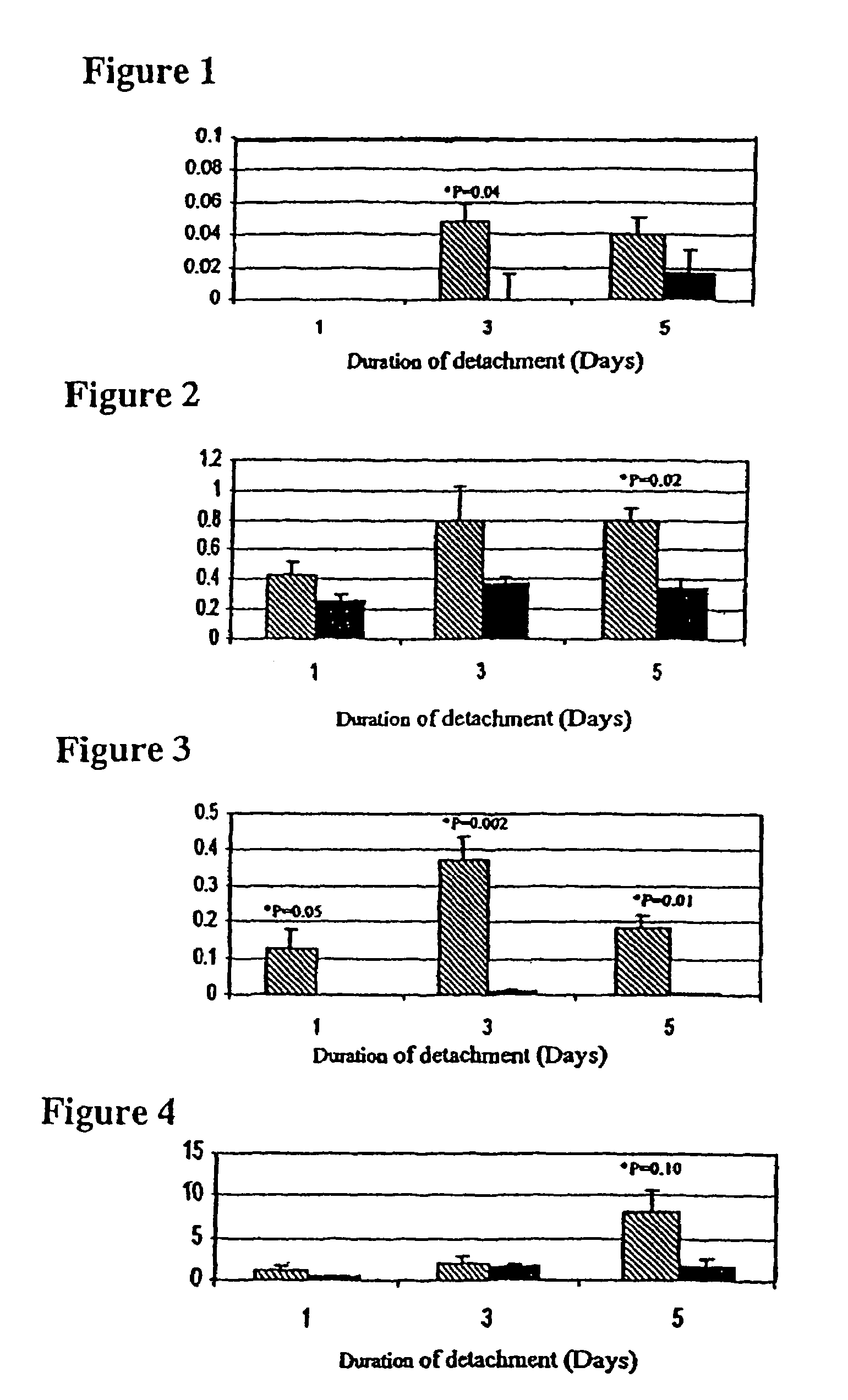
Detection of Caspase Activity Following Retinal Detachment
[0074]This example demonstrates that certain caspases, particularly caspases 3, 7 and 9, are activated in photoreceptor cells following retinal detachment.
[0075]Experimental retinal detachments were created using modifications of previously published protocols (Cook et al. (1995) INVEST. OPHTHALMOL. VIS. SCI. 36(6):990-6; Hisatomi et al. (2001) AM. J. PATH. 158(4):1271-8). Briefly, rats were anesthetized using a 50:50 mixture of ketamine (100 mg / ml) and xylazine (20 mg / ml). Pupils were dilated using a topically applied mixture of phenylephrine (5.0%) and tropicamide (0.8%). A 20 gauge micro-vitreoretinal blade was used to create a sclerotomy approximately 2 mm posterior to the limbus. Care was taken not to damage the lens during the sclerotomy procedure. A Glaser subretinal injector (20 gauge shaft with a 32 gauge tip, Becton-Dickinson, Franklin Lakes, N.J.) connected to a syringe filled with 10 mg / ml of Healon® sodium hyalur...
example 2
Preservation of Photoreceptor Viability Following Retinal Detachment
[0087]The type of experiment provided herein may show that the viability of photoreceptor cells in a detached region of a retina can be maintained by administering a caspase inhibitor to an affected eye.
[0088]Retinal detachments are surgically induced in Brown-Norway rats as discussed in Example 1. The caspase inhibitor, Z-Val-Ala-Asp-fluoromethylketone is dissolved in dimethyl sulfoxide (DMSO) to give the final concentrations of 0.2 mM, 2 mM, and 20 mM. After creating the retinotomy with the subretinal injector, a small amount of Healon® sodium hyaluronate is injected in the subretinal space so as to elevate the retina. After retinal elevation, a Hamilton syringe with a 33 gauge needle is introduced through the retinotomy site, and 25 μl of inhibitor is injected into the region of detachment. About 25 minutes later, Healon® sodium hyaluronate is injected, via the same retinotomy site, to maintain the retinal detach...
example 3
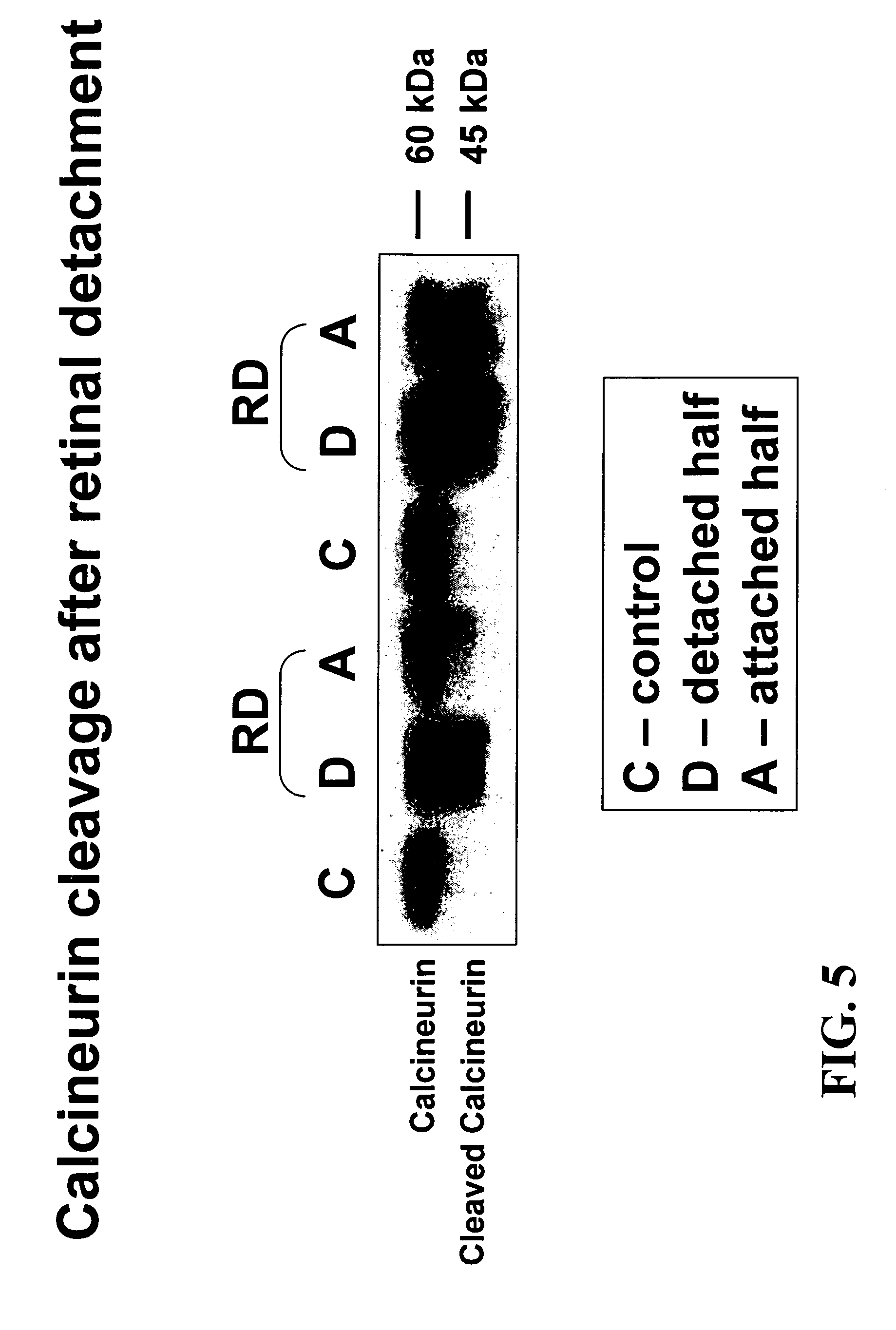
Calcineurin is Cleaved During Retinal Detachment
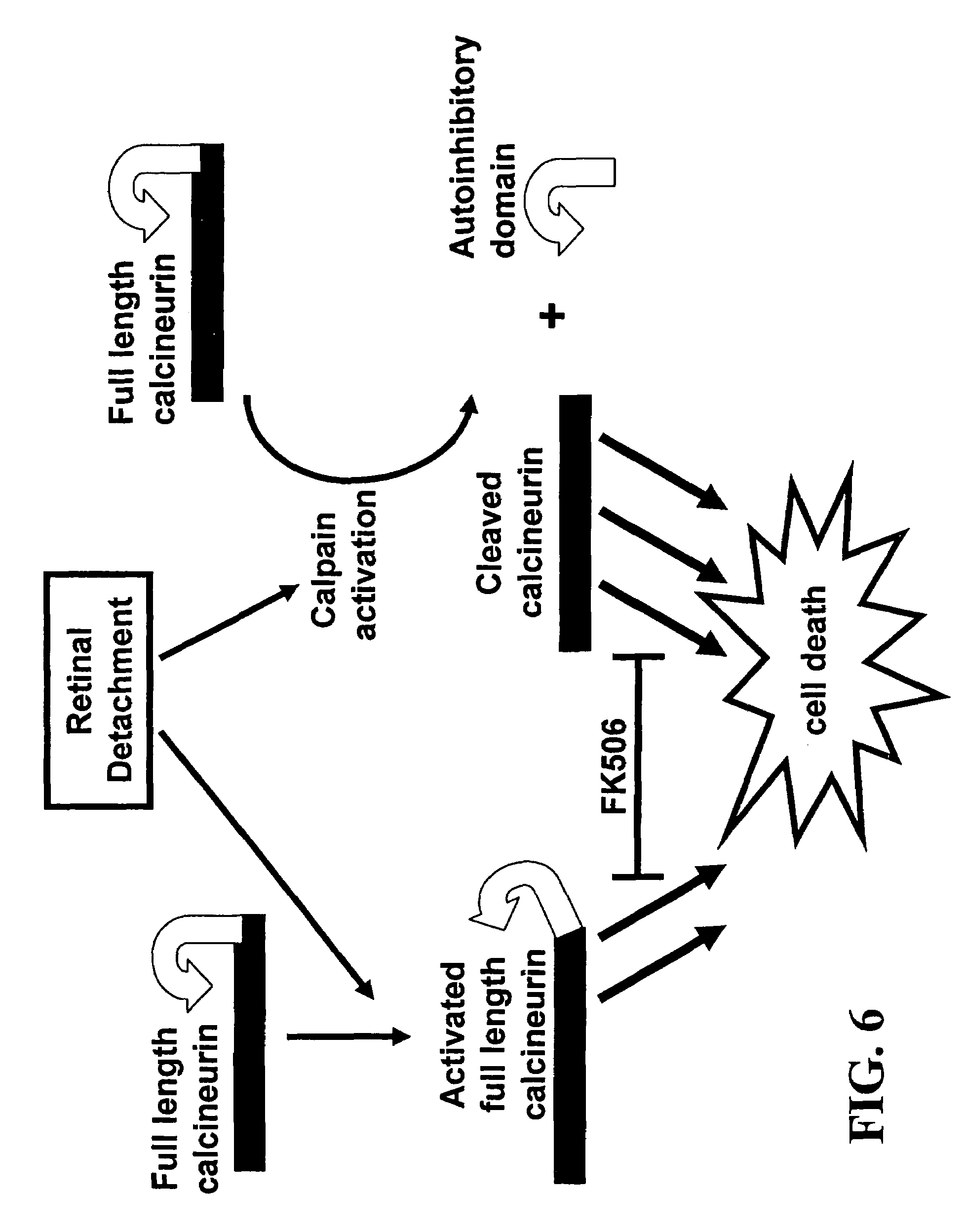
[0091]This experiment provides evidence that in a retinal detachment animal model, calcineurin is cleaved to produce a constitutively active calcineurin molecule (i.e., lacking its autoinhibitory domain). This constitutively active molecule has its active site exposed, but, because it lacks the autoihibitory domain, the active site cannot be hidden. Thus, this cleaved form of calcineurin can activate the apoptotic pathway in photoreceptor cells.
[0092]All experiments were performed in accordance with the ARVO Statement for the Use of Animals in Ophthalmic and Vision Research and the guidelines established by the Animal Care Committee of the Massachusetts Eye and Ear Infirmary. Adult male Brown Norway rats (300-450 g, Charles River, Boston, Mass.) were used in this study, and retinal detachments were created as previously described (Zacks et al. (2003) INVEST. OPHTHALMOL. VIS. SCI. 44(3):1262-7). Rats were anesthetized using a mixture of...
PUM
| Property | Measurement | Unit |
|---|---|---|
| time | aaaaa | aaaaa |
| time | aaaaa | aaaaa |
| time | aaaaa | aaaaa |
Abstract
Description
Claims
Application Information
 - R&D
- Intellectual Property
- Life Sciences
- Materials
- Tech Scout
- Unparalleled Data Quality
- Higher Quality Content
- 60% Fewer Hallucinations
Browse by: Latest US Patents, China's latest patents, Technical Efficacy Thesaurus, Application Domain, Technology Topic, Popular Technical Reports.
© 2025 PatSnap. All rights reserved.Legal|Privacy policy|Modern Slavery Act Transparency Statement|Sitemap|About US| Contact US: help@patsnap.com


