
Apparatus and methods for electrosurgical ablation and resection of target tissue

a target tissue and electrosurgical technology, applied in the field of electrosurgical ablation and target tissue resection, can solve the problems of inability to define the path, damage to or destroy the tissue along the path and surrounding such pathways, and suffer from electrosurgical devices and procedures, etc., and achieve the effect of little or no damag

Inactive Publication Date: 2008-09-30
ARTHROCARE
View PDF423 Cites 217 Cited by
  • Summary
  • Abstract
  • Description
  • Claims
  • Application Information

AI Technical Summary

Benefits of technology

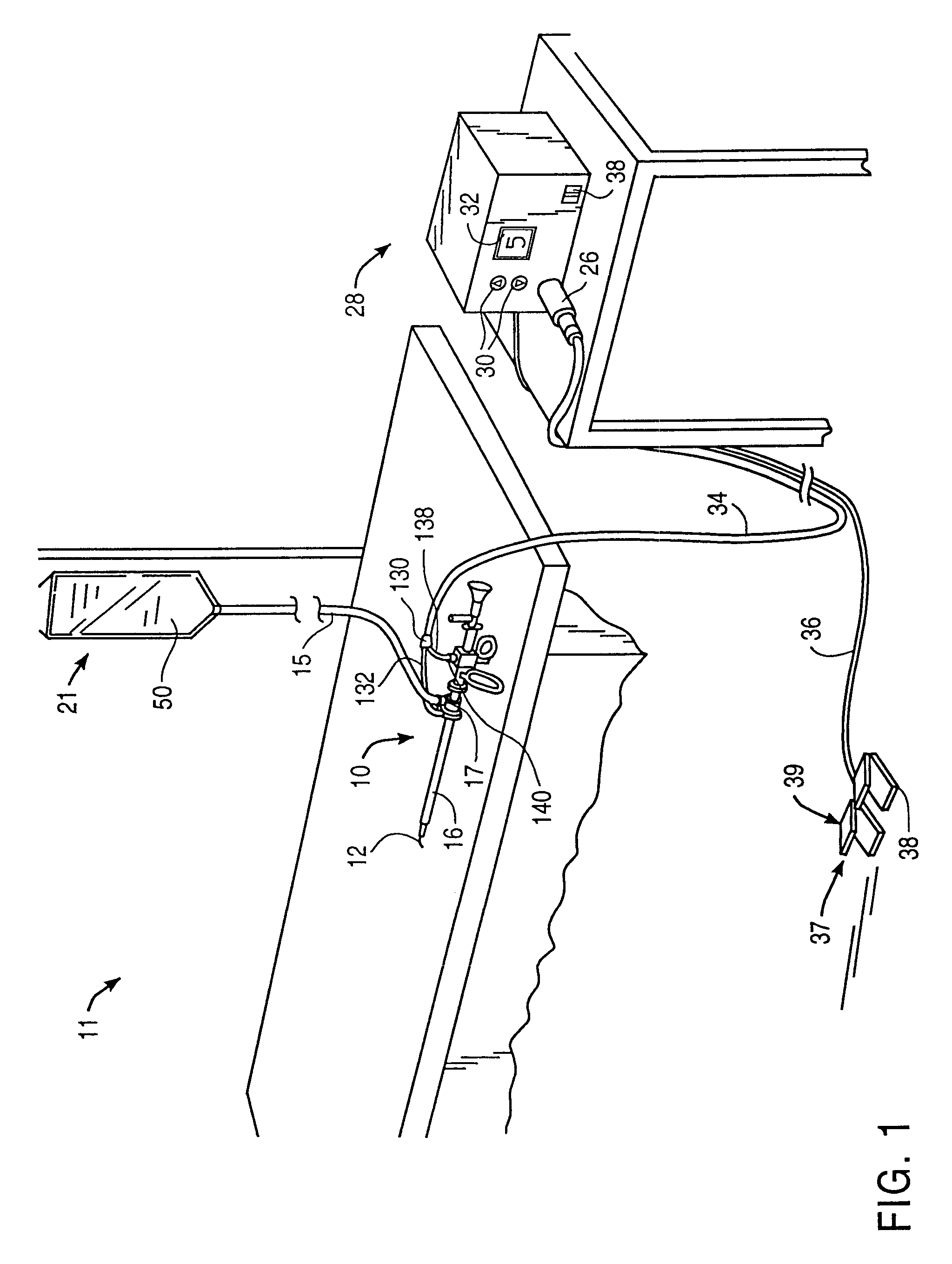
[0011]The present invention provides a system and method for selectively applying electrical energy to structures within or on the surface of a patient's body. The system and method allow the surgical team to perform electrosurgical interventions, such as ablation and cutting of body structures, with little or no damage to non-target tissue adjacent the treatment site. The system and method of the present invention are particularly useful for surgical procedures within accessible sites of the body that are suitable for electrode loop resection, such as the resection of prostate tissue (e.g., transurethral resection of the prostate (TURP)) and leiomyomas (fibroids) located within the uterus.
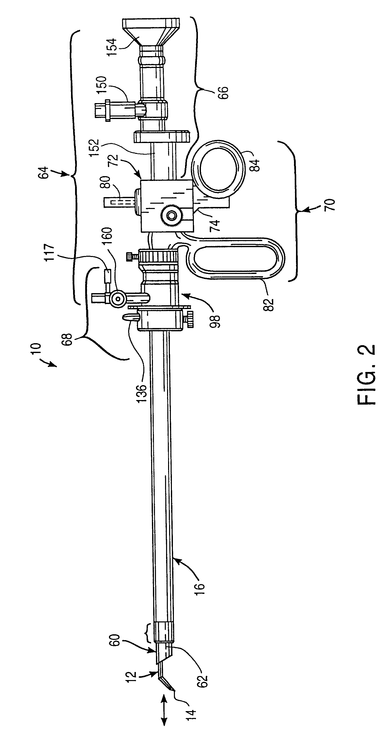
[0012]A system according to one aspect of the present invention comprises an electrosurgical probe having a shaft with a proximal end, a distal end, and at least one active electrode at or near the distal end. A connector may be provided at the proximal end of the shaft for electrically coupling the active electrode to a high frequency voltage source. The active electrode includes at least one active portion having a surface geometry configured to promote substantially high electric field intensities and associated current densities between the active portion and the target site when a high frequency voltage is applied to the electrodes. These high electric field intensities and current densities are sufficient to break down the tissue by processes including molecular dissociation or disintegration of tissue components. The high frequency voltage imparts energy to the target site to ablate a layer of tissue without causing substantial tissue necrosis beyond the boundary of the layer of tissue ablated. This ablative process can be precisely controlled to effect the volumetric removal of tissue as thin as a few layers of cells with minimal or no damage to surrounding or underlying tissue structures.

Problems solved by technology

These electrosurgical devices and procedures, however, suffer from a number of disadvantages.
This current, however, may inadvertently flow along localized, non-defined pathways in the body having less impedance than the defined electrical path.
This situation will substantially increase the current flowing through these non-defined paths, possibly causing damage to or destroying tissue along and surrounding such pathways.
Another limitation of conventional bipolar and monopolar electrosurgery devices is that they are not suitable for the precise removal (ablation) of tissue.
At the point of contact of the electric arcs with tissue, rapid tissue heating occurs due to high current density between the electrode and tissue.
The tissue is parted along the pathway of vaporized cellular fluid, inducing undesirable collateral tissue damage in regions surrounding the target tissue site.
In addition, conventional electrosurgical methods are generally ineffective for ablating certain types of tissue, and in certain types of environments within the body.
For example, loose or elastic connective tissue, such as the synovial tissue in joints, is extremely difficult (if not impossible) to remove with conventional electrosurgical instruments because the flexible tissue tends to move away from the instrument when it is brought against this tissue.
Since conventional techniques rely mainly on conducting current through the tissue, they are not effective when the instrument cannot be brought adjacent to or in contact with the elastic tissue for a long enough period of time to energize the electrode and conduct current through the tissue.
Despite these advantages, laser devices suffer from their own set of deficiencies.
Firstly, laser equipment can be very expensive because of the costs associated with the laser light sources.
Moreover, those lasers which permit acceptable depths of necrosis (such as eximer lasers, erbium:YAG lasers, and the like) provide a very low volumetric ablation rate, which is a particular disadvantage in cutting and ablation of fibrocartilage, articular cartilage, and meniscal tissue.
The holmium:YAG and Nd:YAG lasers provide much higher volumetric ablation rates, but are much less able to control depth of necrosis than are the slower laser devices.
The CO2 lasers provide high rates of ablation and low depth of tissue necrosis, but cannot operate in a liquid-filled cavity.
However, electrolyte-free media have the disadvantage of potentially causing adverse physiological effects in the form of water intoxication, for example, if glycine solution enters the circulation via a severed vein.

Method used


Image

  • Apparatus and methods for electrosurgical ablation and resection of target tissue
  • Apparatus and methods for electrosurgical ablation and resection of target tissue
  • Apparatus and methods for electrosurgical ablation and resection of target tissue

Examples


Embodiment Construction

[0070]The present invention provides a system and method for selectively applying electrical energy to a target location within or on a patient's body, such as solid tissue or the like, including procedures within confined spaces such as the spaces around the articular cartilage between the femur and tibia and spaces between adjacent vertebrae in the patient's spine, and procedures that involve resection of relatively larger pieces of tissue. For convenience, the remaining disclosure will be directed primarily to the resection of prostate tissue, and the cutting, shaping or ablation of meniscal tissue located adjacent articular cartilage and soft tissue covering vertebrae. However, it will be appreciated that the system and method can be applied equally well to procedures involving other tissues of the body, as well as to other procedures including open surgery, laparoscopic surgery, thoracoscopic surgery, and other endoscopic surgical procedures. Examples of such procedures include...


PUM

PropertyMeasurementUnit
distanceaaaaaaaaaa
angleaaaaaaaaaa
angleaaaaaaaaaa

Abstract

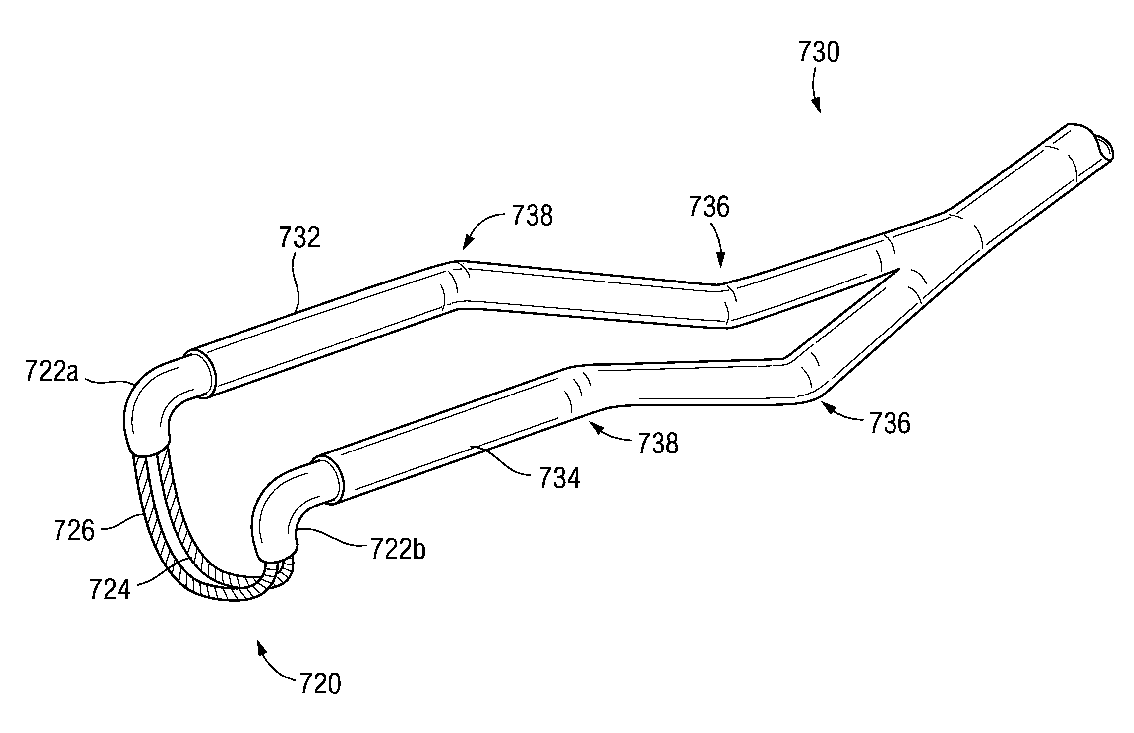
An electrosurgical system and method for ablating, resecting, or cutting body structures, with minimal or no damage to tissue adjacent to the treatment site. The system includes an electrosurgical probe having a shaft with a shaft distal end bifurcated to provide first and second arms. First and second electrode supports are disposed on the first and second arms, respectively. At least one active electrode, in the form of a loop or partial loop, is arranged between the first and second electrode supports. A return electrode, also in the form of a loop or partial loop, is arranged between the first and second electrode supports distal to the active electrode. The active and return electrodes are configured to promote substantially high electric field intensities and associated high current densities between the active portion and the target site when a high frequency voltage is applied to the electrodes. These high current densities are sufficient to break down the tissue by processes including molecular dissociation of tissue components. In one embodiment, the high frequency voltage imparts energy to the target site to effect the vaporization and volumetric removal of a layer of tissue without causing substantial tissue damage beyond the layer of tissue ablated. In another embodiment, a fragment of target tissue is removed, with minimal or no damage to surrounding tissue, by a process including the molecular dissociation of tissue components, and the tissue fragment is retrieved for biopsy.

Description

CROSS-REFERENCE TO RELATED APPLICATIONS[0001]The present invention is a continuation-in-part of U.S. patent application Ser. No. 09 / 571,343, filed May 16, 2000 now U.S. Pat. No. 6,575,968, which is a Divisional of U.S. patent application Ser. No. 08 / 687,792, filed Jul. 18, 1996, now U.S. Pat. No. 5,843,019, which is a continuation-in-part of application Ser. No. 08 / 561,958, filed on Nov. 22, 1995, now U.S. Pat. No. 5,697,882, which is a continuation-in-part of application Ser. No. 08 / 485,219, filed on Jun. 7, 1995, now U.S. Pat. No. 5,697,281, which is a continuation-in-part of PCT International Application, U.S. National Phase Ser. No. PCT / US94 / 05168, filed on May 10, 1994, which was a continuation-in-part of application Ser. No. 08 / 059,681, filed on May 10, 1993 now abandoned, which is a continuation-in-part of application Ser. No. 07 / 958,977, filed on Oct. 9, 1992, now U.S. Pat. No. 5,366,443, which is a continuation-in-part of application Ser. No. 07 / 817,575, filed on Jan. 7, 19...

Claims


Application Information

Patent Timeline
Patent Type & Authority Patents(United States)
IPC IPC(8): A61B18/18A61B17/00A61B17/22A61B18/00A61B18/12A61B18/14A61B19/00A61F2/02A61M3/02A61M25/01
CPCA61B18/12A61B18/1402A61B18/148A61B18/1482A61B18/1485A61B18/149A61B18/1492A61M25/0133A61B18/1206A61M25/0158A61B2017/00026A61B2017/00084A61B2017/00097A61B2017/00101A61B2017/00247A61B2017/00292A61B2017/003A61B2017/22001A61B2017/22038A61B2018/00029A61B2018/00083A61B2018/00119A61B2018/0016A61B2018/00178A61B2018/00196A61B2018/00327A61B2018/00392A61B2018/00505A61B2018/00577A61B2018/00583A61B2018/00642A61B2018/0066A61B2018/00678A61B2018/00702A61B2018/00726A61B2018/00761A61B2018/00791A61B2018/00797A61B2018/00821A61B2018/00827A61B2018/00875A61B2018/00886A61B2018/1213A61B2018/124A61B2018/1253A61B2018/126A61B2018/1273A61B2018/1467A61B2018/1472A61B2018/1497A61B2018/162A61B2018/165A61B2019/5276A61B2218/002A61F2/2493A61M3/0283A61M25/0147A61B2090/378
Inventor WOLOSZKO, JEANDAVISON, PAULEGGERS, PHILIP E.THAPLIYAL, HIRA V.
Owner ARTHROCARE
Learn More