
Intraoperative neurophysiological monitoring system

a neurophysiological monitoring and intraoperative technology, applied in the field of surgical instruments, can solve the problems of significant risk of facial nerve loss following head and neck surgery such as acoustic neuroma resection, nerves are very delicate, and there is a considerable risk of nerve bruise, stretch or severe injury during operation, so as to improve the sensitivity of recording electrical artifacts

Active Publication Date: 2007-05-08
PRASS RICHARD L
View PDF68 Cites 167 Cited by
  • Summary
  • Abstract
  • Description
  • Claims
  • Application Information

AI Technical Summary

Benefits of technology

"The present invention is an intraoperative neuropathological monitoring system that uses a digitally controlled stimulator with multiple independent stimulus outputs, an artifact detection electrode with modified wire leads, a method and algorithm for detecting brief artifacts, a method and algorithm for controlling the sequence of monitoring events, and a method and algorithm for using the stimulator probe as an input device. The system allows for the safe and effective monitoring of nerve integrity during surgery, minimizing stimulation-related recording artifacts, and improving the accuracy and safety of stimulus delivery. The system also includes a mechanism for detecting the duration of continuous contact of the stimulus probe with patient tissue, which triggers a compensatory increase in delivered stimulus current to prevent stimulus shunting. Overall, the invention provides a more efficient and effective means for monitoring nerve integrity during surgery."

Problems solved by technology

Despite advances in diagnosis, microsurgical techniques, and neurotological techniques enabling more positive anatomical identification of facial nerves, loss of facial nerve function following head and neck surgery such as acoustic neuroma resection is a significant risk.
Nerves are very delicate and even the best and most experienced surgeons, using the most sophisticated equipment known, encounter a considerable hazard that a nerve will be bruised, stretched or severed during an operation.
Prior art nerve integrity monitoring instruments (such as the Xomed® NIM-2® XL Nerve Integrity Monitor, manufactured by the assignee of the present invention) have proven to be effective for performing the basic functions associated with nerve integrity monitoring such as recording EMG activity from muscles innervated by an affected nerve and alerting a surgeon when the affected nerve is activated by application of a stimulus signal, but have significant limitations for some surgical applications and in some operating room environments.
A first problem is users have noticed certain EMG measurement artifacts have a disruptive effect on monitoring and tend to cause undesirable false alarms.
In particular, EMG monitoring often is performed during electrocautery in a surgical procedure, wherein powerful currents surge through and cauterize the tissue, often to devastating effect on the monitor's sensitive amplifier circuits.
These brief artifacts may be confused for true electromyographic (muscle) responses and may lead to misinterpretation and false alarms, thereby reducing user confidence and satisfaction in nerve integrity monitoring.
Maintenance of high common-mode rejection characteristics in the signal conditioning path has helped to reduce such interference, however, false alarms still occur.
Prior art nerve integrity monitoring devices incorporate a simple threshold detection method to identify significant electrical events based upon the amplitude of the signal voltage observed in the monitoring electrodes, relative to a baseline of electrical silence, a methodology having disadvantages for intraoperative nerve integrity monitoring.
Moreover, with close electrode spacing and bipolar amplification, recording of larger responses is frequently associated with internal signal cancellation, significantly reducing the amplitude of the observed electrical signal.
Another disadvantage of prior art methodology of threshold detection is that the surgeon cannot readily distinguish or select between electrical artifacts and EMG activity.
A second problem is that the nerves of interest may frequently exhibit a variable amount of irritability during the surgical procedure, which may be caused by a disease process or by surgical manipulations such as mild traction or by drying or thermal effects.
Once a reasonable effort to reduce nerve irritability has been carried out, any residual nerve irritability becomes “noise” and may interfere with the ability to detect electrically and mechanically stimulated nerve activity.
A third problem arises when monopolar probes, bipolar probes or electrified instruments are selected for electrical stimulation during intraoperative neurophysiological monitoring.
If there are two outputs, the outputs are connected in parallel with a single common stimulus intensity setting and so there is no ability to provide separate (optimized) stimulus intensities or to guard against parallel communication between the two outputs.
If both outputs are connected to stimulus instruments, undetected current-leak could occur through parallel channels and result in false-negative stimulation.
At least one manufacturer or prior art monitoring instruments offers a switchable connector at the stimulus probe terminus, allowing more than one stimulus instrument to be kept in readiness, and avoiding parallel connections to the unused instruments, but performing the act of switching requires a surgical staff member such as a nurse or technician and so is cumbersome and, being time consuming, expensive.
A related problem is that prolonged nerve irritability may be due to light anesthesia, rather than to inherent nerve irritability.
Another problem confronting users of prior art nerve integrity monitoring devices is that quantative measurements of nerve function are relatively cumbersome to obtain, since equipment setting changes must be performed by operating room personnel while electrical stimulation procedures are performed by the operating surgeon.
With prior art technology, this process is time-intensive and discourages serial determinations during the operation as an ongoing measure of nerve integrity.
When using prior art methods, if the threshold is found to be abnormal, the surgeon is usually unaware of when the change to abnormality occurred during the operative procedure.
For multi-channel nerve integrity monitoring with qualitative and quantitative signal analysis, front and back panel hardware is cumbersome and too limited in scope.
A limitation of prior art strategies is that the setup information is held in volatile memory during actual monitoring operations, rendering the setup information vulnerable to strong electrical surges, electromagnetic noise or accidental power interruptions.
An electrical surge or accidental unplugging may cause loss of all new (different from “default”) setup information, requiring a “reboot” of the system and adjustment to get back to the desired settings.
Stimulation devices of the prior art for neurophysiological monitoring are manually controlled through front panel potentiometers and switches or with mouse and keyboard to produce paired or burst stimuli and stimuli of opposite polarity in an alternating pattern, but lack the ability to deliver consecutive stimuli of differing intensities or alter the pattern of stimulation at a predetermined time without that time consuming manual input.
Analogously, none of the monitoring instruments of the prior art provide delivery of selected stimuli in coordination with data acquisition, analysis, display, and storage.
Moreover, in prior art nerve integrity devices, control of on-line functions is performed by keyboard and mouse or by front panel controls and, because of a possible breach of sterility, the operating surgeon cannot perform such functions by himself or herself and so changing equipment settings requires involvement of hospital personnel at the request of the operating surgeon and may be time-consuming, cumbersome and possibly risky, since the changed settings may be inaccurate.
Such irritability may be due to nerve compromise caused by the disease process itself or to various surgical manipulations, such as mild traction, drying, thermal stimulation, or chemical irritation.
Once a reasonable attempt has been made to allow the nerve to become quieted, any remaining repetitive activity is essentially “noise” and may interfere with hearing more important brief EMG responses that allow localization of the nerve of interest.
Such background irritability is particularly a problem during acoustic neuroma resections, which is one of the most common procedures for which facial nerve monitoring is used.
The majority of prior art nerve integrity monitoring devices have only two channels, which allows little redundancy and flexibility.
When repetitive activity becomes bothersome and persistent, despite reasonable efforts on the part of the operating surgeon to allow the nerve to quiet down, the surgeon may ask an operating room employee to “turn the monitor down.” This solution is problematic, because it may cause the surgeon to miss hearing important localizing EMG information.
However, even with visual displays, the process may still be time consuming and, therefore, expensive.
Moreover, once certain “offending” channels have been muted, there may be long periods before the surgeon, the nurse, or operating room technician remember or “feel safe” to add these channels back to the audio signal.
This may cause unnecessarily long periods of decreased sensitivity.

Method used


Image

  • Intraoperative neurophysiological monitoring system
  • Intraoperative neurophysiological monitoring system
  • Intraoperative neurophysiological monitoring system

Examples


Embodiment Construction

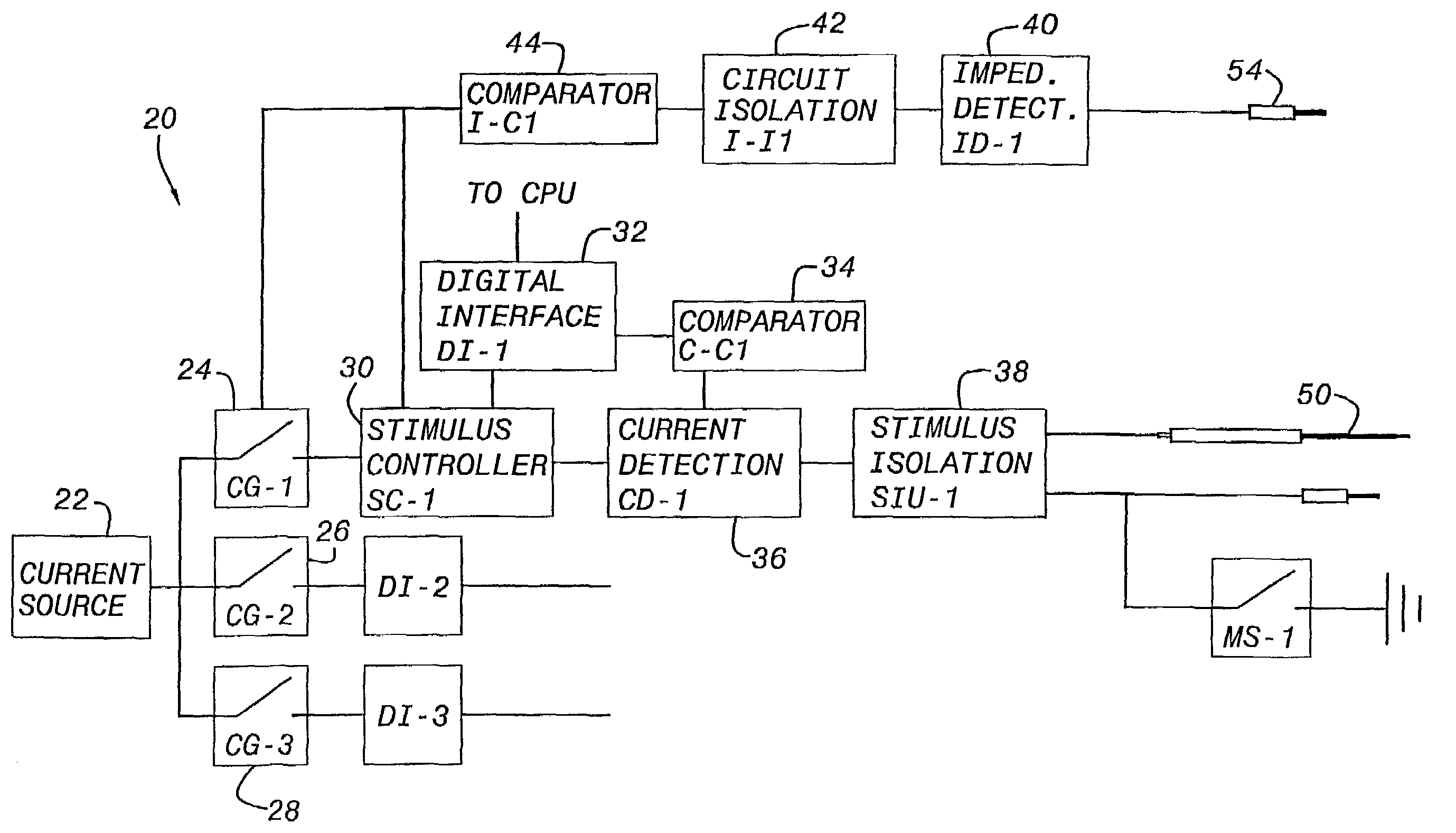
[0100]Referring specifically to FIG. 1 of the accompanying drawings, an intraoperative neurophysiological monitoring system 20 includes digitally controlled stimulator impedance and current flow detection circuit elements for use during intraoperative neurophysiological monitoring and preferably in conjunction with a tissue contact initiated event sequencing time line algorithm (TCI-Time line) for control of data acquisition, analysis, display and storage. A current source 20 is connected with parallel inputs to three electronic switches (“current-gates”) CG-1, CG-2, and CG-3, 24, 26 and 28; although separate current sources for each stimulus output may be employed, a single current source is shown here. Each stimulus output includes a stimulus-controller (e.g., SC-1) 30 controlling the intensity, duration and temporal patterns of delivered stimulus-pulses. The controller is, in turn, connected with controlled by and responsive to a digital-interface (e.g., DI-1) 32 which is in turn...


PUM


Abstract

An intraoperative neurophysiological monitoring system includes an adaptive threshold detection circuit adapted for use in monitoring with a plurality of electrodes placed in muscles which are enervated by a selected nerve and muscles not enervated by the nerve. Nerve monitoring controller algorithms permit the rapid and reliable discrimination between non-repetitive electromyographic (EMG) events repetitive EMG events, thus allowing the surgeon to evaluate whether nerve fatigue is rendering the monitoring results less reliable and whether anesthesia is wearing off. The intraoperative monitoring system is designed as a “surgeon's monitor,” and does not require a neurophysiologist or technician to be in attendance during surgery. The advanced features of the intraoperative monitoring system will greatly assist neurophysiological research toward the general advancement of the field intraoperative EMG monitoring through post-surgical analysis. The intraoperative monitoring system is preferably modular, in order to allow for differential system pricing and upgrading as well as to allow for advances in computer technology; modularity can also aid in execution of the design.

Description

BACKGROUND OF THE INVENTION[0001]1. Field of the Invention[0002]The present invention relates to surgical apparatus and more particularly to a neurophysiological monitoring system including a nerve integrity monitoring instrument for use in conjunction with one or more electrical stimulus probes as an intraoperative aid in defining the course of neural structures. The invention is particularly applicable for use in monitoring facial electromyographic (EMG) activity during surgeries in which a facial motor nerve is at risk due to unintentional manipulation, although it will be appreciated that the invention has broader applications and can be used in other neural monitoring procedures.[0003]2. Discussion of the Prior Art[0004]Despite advances in diagnosis, microsurgical techniques, and neurotological techniques enabling more positive anatomical identification of facial nerves, loss of facial nerve function following head and neck surgery such as acoustic neuroma resection is a signif...

Claims


Application Information

Patent Timeline
Patent Type & Authority Patents(United States)
IPC IPC(8): A61B5/05A61B5/04A61N1/00A61B5/0488A61B5/053A61N1/05A61N1/08
CPCA61B5/0488A61B5/0531A61B5/4821A61B5/4893A61B5/6843A61N1/08A61B5/7217A61B5/389A61B5/395
Inventor PRASS, RICHARD L.
Owner PRASS RICHARD L
Learn More