
Tumor specific monoclonal antibodies

a monoclonal antibody and tumor-specific technology, applied in the field of cancer, can solve the problems of lack of malignant tumor development, and inability to achieve specificity for cancer cells, and achieve the effect of reducing the proliferation of neoplastic cells

Inactive Publication Date: 2006-10-03
APPLIED MOLECULAR EVOLUTION
View PDF1 Cites 35 Cited by
  • Summary
  • Abstract
  • Description
  • Claims
  • Application Information

AI Technical Summary

Benefits of technology

[0012]A method for reducing neoplastic cell proliferation is also provided. The method consists of administering an effective amount of a human monoclonal antibody or functional fragment of the invention. Also provided is a method of detecting a neoplastic cell in a sample. The method consists of contacting a cell with a mono

Problems solved by technology

In cancer, neoplastic cells escape from their normal growth regulatory mechanisms and proliferate in an uncontrolled fashion, leading to the development of a malignant tumor.
A major problem with each of these treatments is their lack of specificity for cancer cells.
For instance, surgical removal of the tumor is often incomplete.
Even a few residual neoplastic cells can be lethal, as they can rapidly proliferate and metastasize to other sites.
Radiation and chemotherapy also have serious limitations.
Due to their toxicity to normal tissues, the amount of radiation or chemotherapeutic agent that can be safely used is often inadequate to kill all neoplastic cells.
Additionally, their toxicity to normal tissues is manifested by unpleasant side effects, including nausea and hair loss, that severely reduce the quality of life for the cancer patient undergoing these treatments.
Most current immunotherapeutic strategies for cancer have been of limited utility due to their reliance on mouse monoclonal antibodies.
Thus, human tumor-specific antigens often fail to generate therapeutically beneficial murine antibodies.
Furthermore, the human immune system is less likely to generate antigens against epitopes present on normal cells, increasing the odds of generating and successfully identifying tumor-specific antigens.
These procedures are seriously limited by the narrow range of antigen specificities of activated B-cells of cancer patients.
As it is clearly not possible to immunize individuals in vivo with tumor cells, as one can with mice, it has not been possible to readily generate tumor-specific human monoclonal antibodies to any given antigen or tumor cell type.

Method used


Image

  • Tumor specific monoclonal antibodies
  • Tumor specific monoclonal antibodies
  • Tumor specific monoclonal antibodies

Examples


example i

Production of Tumor-Specific Human Monoclonal Antibodies

[0149]This Example shows production of hybridomas that secrete human monoclonal antibodies specifically reactive with tumor cells. A procedure for immunizing normal lymphocytes in vitro with tumor cells or cell membranes prior to immortalization is described. This procedure allowed enrichment of lymphocytes producing novel tumor-reactive human monoclonal antibodies. These human monoclonal antibodies will be useful for cancer immunotherapeutic and immunodiagnostic procedures.

[0150]Lymphocytes were prepared as follows. Spleen tissue was isolated from accident victims, fragmented, and forced through a no. 50 mesh wire screen (Bellco, Vineland, N.J.). The cells were collected by centrifugation at 250×g for 10 min and RBC were removed by ammonium chloride lysis. The remaining cells were washed, resuspended in freezing medium (40% RPMI, 50% FCS and 10% DMSO) at a concentration of 100 to 300×106 cells / ml, frozen in 1.5-ml aliquots, an...

example ii

Immunoreactivities of LH13 and LH11238 Antibodies

[0175]This Example shows the immunoreactivities of LH13 and LH11238 antibodies with a panel of human tumor cells.

[0176]In order to use LH13 and LH11238 antibodies for immunodiagnostic and immunotherapeutic purposes, the range of tumor types expressing the corresponding antigens needs to be identified. Tumor cell lines are representative of the corresponding tumor type, and normal fibroblasts are representative of non-neoplastic tissues. The tumor cell lines were derived from melanomas, lung carcinomas, ovarian carcinomas and breast carcinomas. The human monoclonal antibodies of the invention were tested for immunoreactivity with both human cancer cell lines and normal fibroblasts.

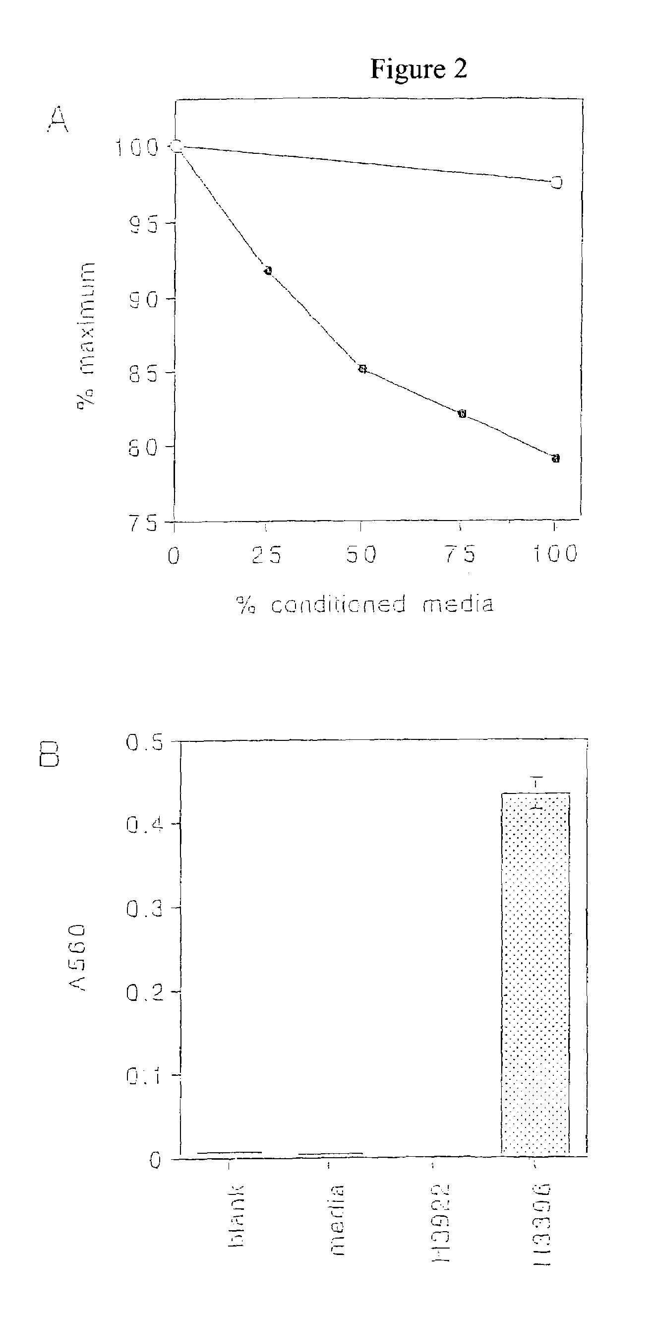
[0177]H3396, H3464, H3477 and H3922 are cultured cell lines established from metastases of human breast adenocarcinoma, which were explanted and maintained in culutre. H2981 and H2987 are cultured cell lines established from human lung carcinomas, which were ...

example iii

Binding Activity of LH13 and LH11238 Human Monoclonal Antibodies

[0182]This Example shows the binding activity of human monoclonal antibodies LH13 and LH11238 with normal and tumor cells.

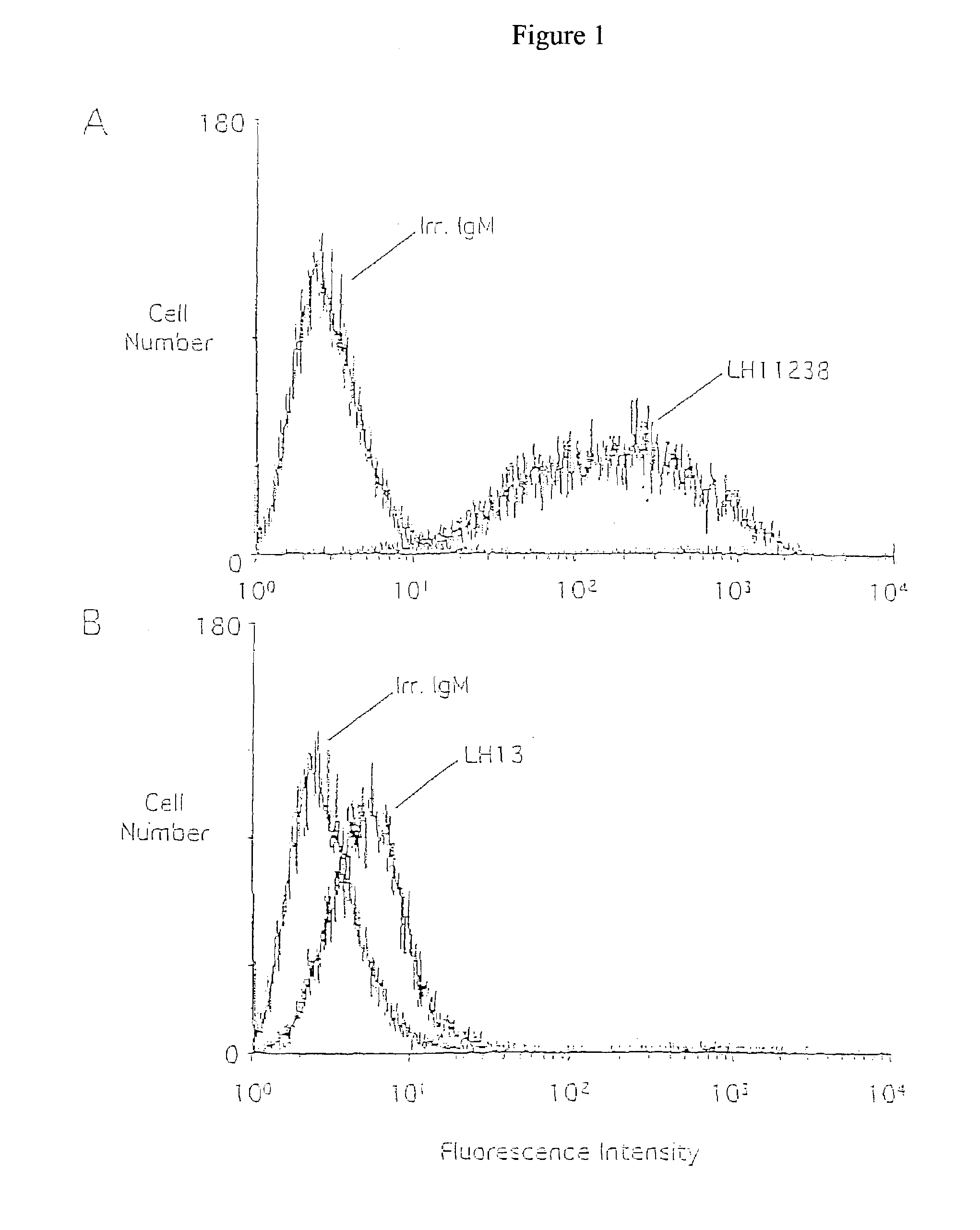
[0183]In order to determine the immunoreactivity of human monoclonal antibodies with live tumor cells and normal cells, flow cytometry (FACS analysis) was used. H3464 breast tumor cells were chosen because they expressed both LH13 and LH11238 antigens by ELISA analysis and were readily isolated as a single cell suspension. Normal peripheral blood lymphocytes were also examined as representative of normal tissues. Facs analysis additionally permits examination of heterogeneity of a cell population with respect to antigen expression.

[0184]H3464 cells were removed from culture dishes with trypsin or Versene (EDTA), washed with PBS, resuspended at 2×10⁶ cells / ml in tumor media, and allowed to recover 2 h at 37° C. Antibodies were incubated at 5.0 μg / ml with 1×10⁶ tumor cells in 50 μl total volume for 30 ...


PUM

PropertyMeasurementUnit
Fractionaaaaaaaaaa
Capacitanceaaaaaaaaaa
Cytotoxicityaaaaaaaaaa

Abstract

The invention provides tumor-specific human monoclonal antibodies and functional fragments. Also provided are nucleic acids encoding tumor-specific human monoclonal antibodies and functional fragments. A method for reducing neoplastic cell proliferation is also provided. The method consists of administering an effective amount of a tumor-specific human monoclonal antibody or functional fragment. Also provided is a method of detecting a neoplastic cell in a sample. The method consists of contacting a cell with a tumor-specific monoclonal antibody or functional fragment and detecting the specific binding of the human monoclonal antibody or functional fragment to the sample.

Description

[0001]This application claims benefit of the filing date of U.S. Provisional Application No. 60 / 421,146 (yet to be assigned), filed Nov. 19, 2001, which was converted from U.S. Ser. No. 09 / 989,901, and which is incorporated herein by reference.BACKGROUND OF THE INVENTION[0002]The present invention relates generally to cancer and, more specifically, to human monoclonal antibodies for the treatment and diagnosis of cancer.[0003]Cancer is one of the leading causes of death in the United States. Each year, more than half a million Americans die from cancer, and more than one million are newly diagnosed with the disease. In cancer, neoplastic cells escape from their normal growth regulatory mechanisms and proliferate in an uncontrolled fashion, leading to the development of a malignant tumor. Tumor cells can metastasize to secondary sites if treatment of the primary tumor is either not complete or not initiated before substantial progression of the disease. Early diagnosis and effective ...

Claims


Application Information

Patent Timeline
IPC IPC(8): C07K16/00C07K16/30
CPCA61K2039/505A61K2039/5152C07K16/00C07K16/3015C07K2317/55C07K2317/565C07K2317/77
Inventor WATKINS, JEFFRY D.
Owner APPLIED MOLECULAR EVOLUTION
Learn More