
Medical device with elastomeric penetrable wall and inner seal

a technology of elastomeric and penetrating walls, which is applied in the direction of medical devices, intravenous devices, medical devices, etc., can solve the problems of difficult swab, cumbersome solutions, and one is forced to pay more for swab

Inactive Publication Date: 2006-06-06
B BRAUN MEDICAL
View PDF67 Cites 81 Cited by
  • Summary
  • Abstract
  • Description
  • Claims
  • Application Information

AI Technical Summary

Benefits of technology

[0013]Locking systems may be used as additional structure for connection, so that additional connector separation can be further eliminated as a risk.
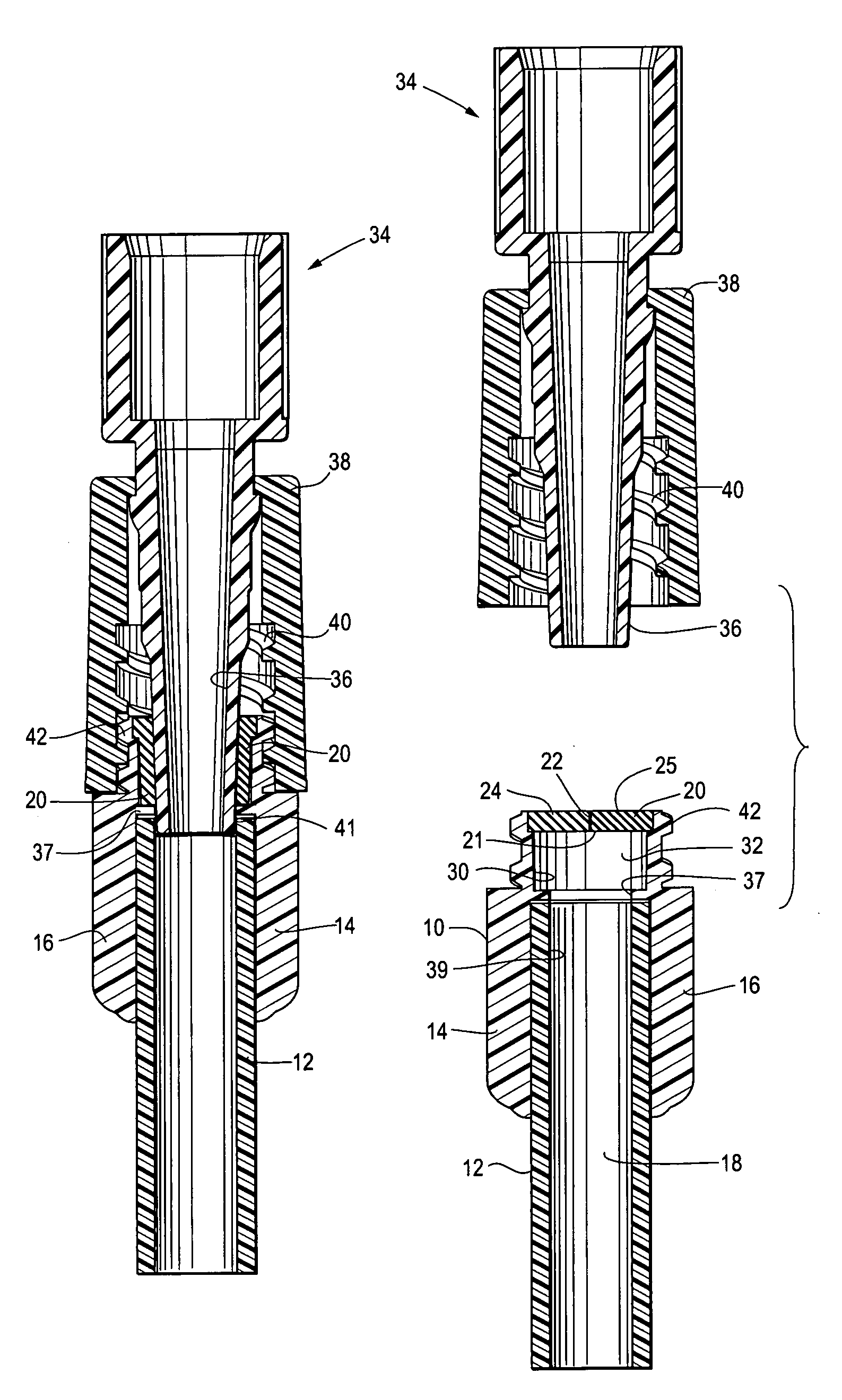
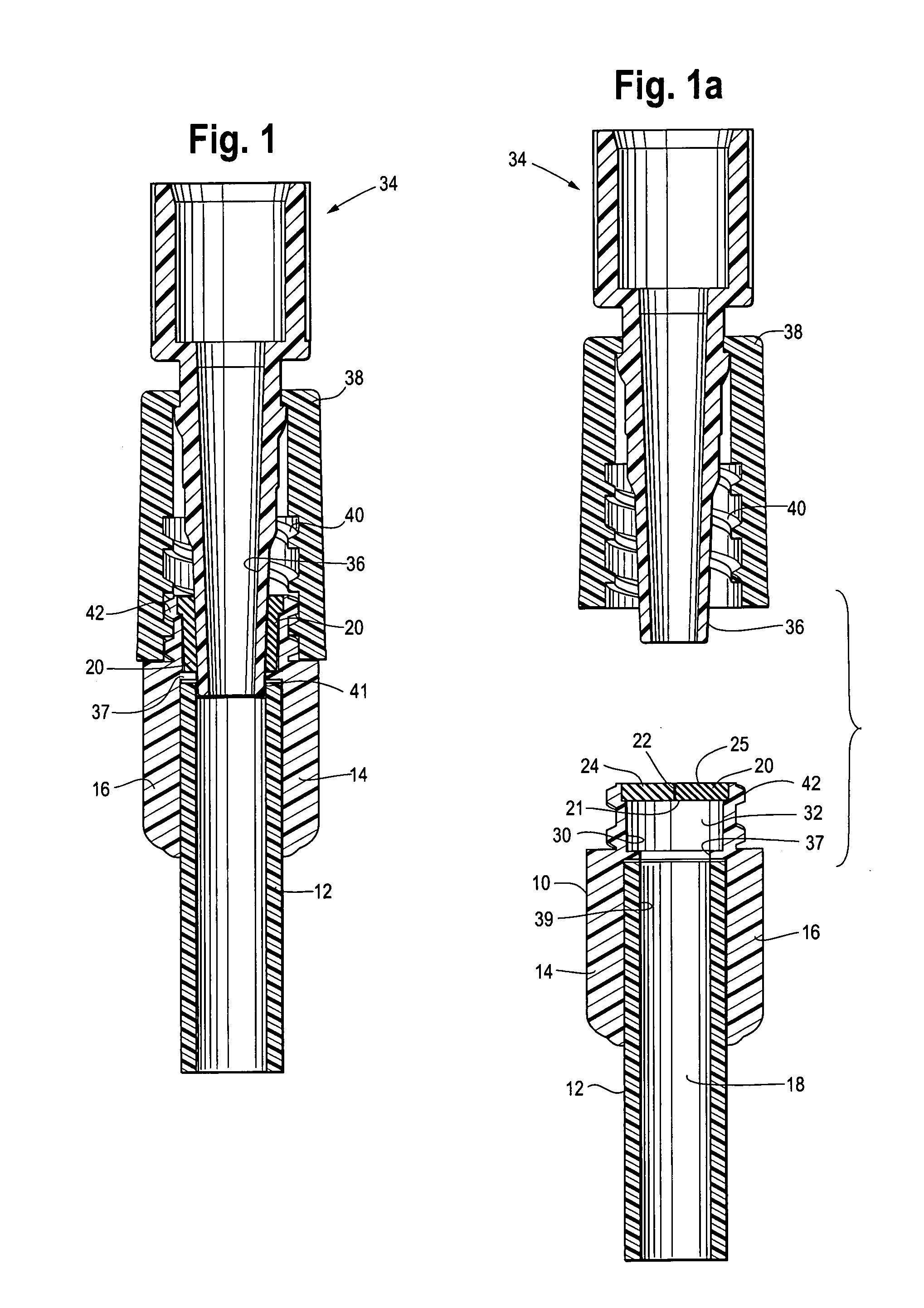
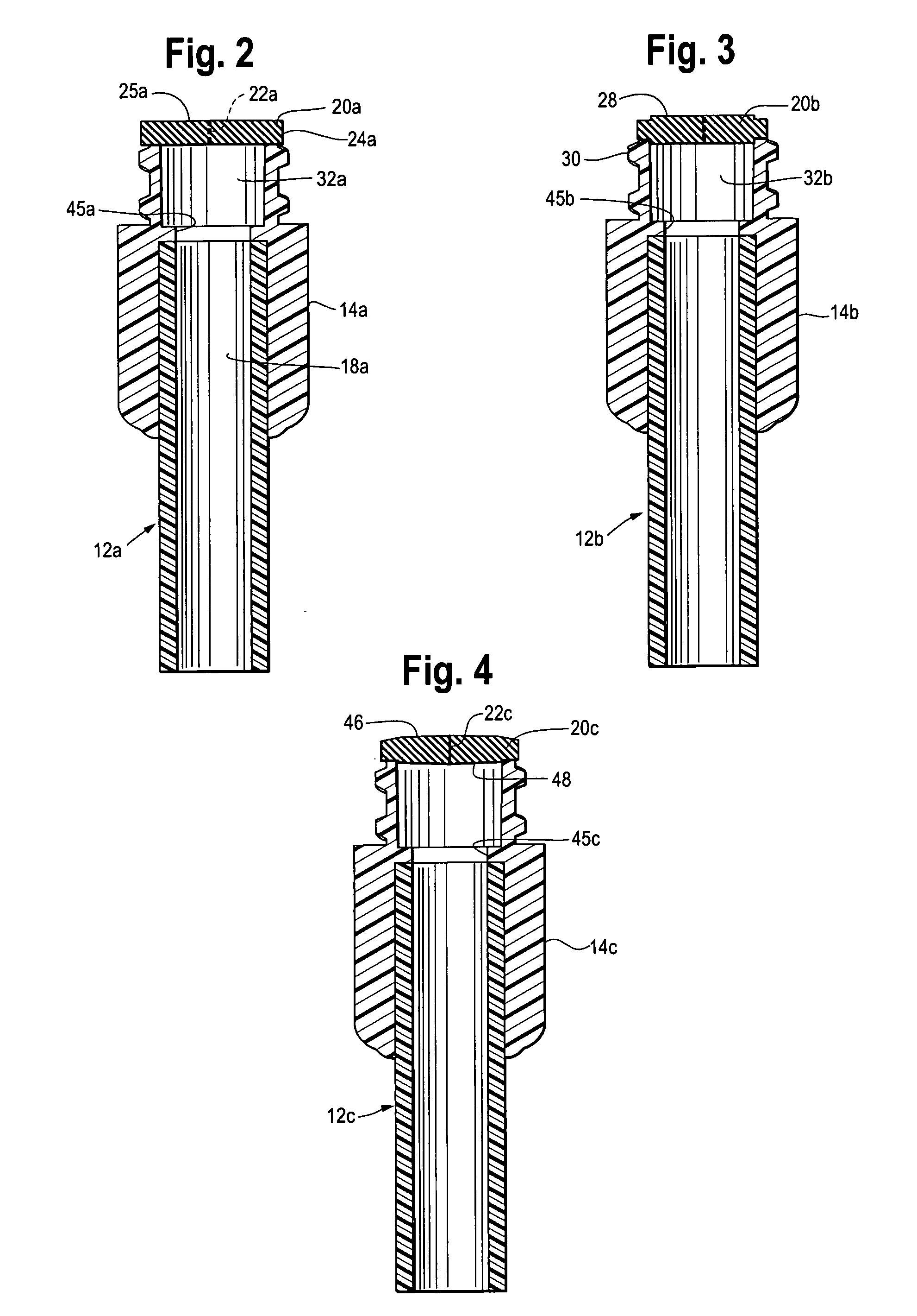
[0016]Thus, one currently preferred aspect of this invention is to shift a seal between the medical device and a male luer or other connector tube from conventional positions that are outside of the elastomeric wall to a generally new position which is within the device and under the elastomeric wall. With such a seal being inside of the elastomeric wall, a simplified elastomeric wall can be used without major, extending skirts or other appendages as in much of the prior art, while such an elastomeric wall can be swabbed for aseptic cleaning, and good, reliable sealing and retention between the male luer and the medical device can still be achieved.
[0019]The device of this invention may define a bore portion positioned between the elastomeric wall and the passageway recited above. This bore portion may have an inner diameter which is larger than any inner diameter of the passageway, to provide an annular chamber that receives the material of the elastomeric wall when a connector tube is extending through the perforation. Then, as stated, the connector tube, typically a male luer, penetrates the perforation and forms an annular seal with the elastomeric wall. Preferably, below said bore portion, which in some embodiments is frustoconical as stated above, the male luer may also create an additional, annular seal with the passageway wall. The connector tube may have a frustoconical outer wall with an angle which substantially matches the frustoconical passageway wall, to facilitate the annular seal. Alternatively, the length and / or strength of the conical bond between the male luer and passageway wall may be reduced, if desired, by building in a slight difference in frustoconical angle between the two parts. Typically the angle in question approximates the angle of an ISO luer system, namely about 1–4° of all sides to the longitudinal axis of the bore.

Problems solved by technology

This in turn raises a problem, in that there is a need for assuring reliable connection of a male luer, for example, residing in the elastomeric wall as it extends therethrough to provide fluid communication.
However, if the elastomeric wall is positioned within such a sleeve, then it is difficult to swab.
If it is exposed so as to be swabbable, then one has, up until now, been forced toward more expensive and cumbersome solutions of the problem.

Method used


Image

  • Medical device with elastomeric penetrable wall and inner seal
  • Medical device with elastomeric penetrable wall and inner seal
  • Medical device with elastomeric penetrable wall and inner seal

Examples


Embodiment Construction

[0006]In accordance with this invention, an injection site is provided as part of a medical device which has an interior for containment of a fluid (liquid or gas). For example, the medical device may be a drug vial or container which utilizes the injection site of this invention, or it can be a tubing set having a main flow path for blood, parenteral solution, gases, or the like, to permit access preferably either by a male luer (luer slip or luer lock) connector or other type of tubular probe, without any intermediate device as in the prior art, such access being to the flow path of the tube set or the interior of any other container. Preferably, access through the injection site is also available as well by a needle, sharp or blunt.

[0007]An opening is defined into the interior of the medical device, with an elastomeric wall forming a fluid / air tight barrier across said opening, preferably both at positive and negative pressures relative to atmosphere. The elastomeric wall typical...


PUM


Abstract

A medical device has an interior for containment of fluids; an opening into the interior, and an elastomeric wall having at least one perforation. The wall comprises a fixedly placed, flexible barrier across the opening. The device also defines a passageway extending inwardly from adjacent to the elastomeric wall, the passageway being proportioned to receive and form an annular seal with a connector tube that extends into the interior through the perforation.

Description

[0001]This is a continuation-in-part of Utterberg, et al. application Ser. No. 10 / 264,863, filed Oct. 4, 2002.BACKGROUND OF THE INVENTION[0002]Tubing sets for the transfer of medical fluids such as blood or parenteral solution generally comprise flexible, plastic tubing with end connectors, which are often luer-type connectors. Injection sites are often carried on the tubing at the ends of, or between the ends of, the tubing sets, comprising an elastomeric diaphragm or bung which is carried in a housing in typically compressed or restrained fashion, so that a needle can resealably penetrate the elastomeric bung and communicate with the interior of the tubular set, for administration of medicines and the withdrawal of medical samples such as blood. Also, in the field of extracorporeal blood transport, in which blood is conveyed to and from an extracorporeal blood processing device such as a dialyzer or an aphaeresis apparatus, such an injection site may also be used to add parenteral...

Claims


Application Information

Patent Timeline
Patent Type & Authority Patents(United States)
IPC IPC(8): A61M5/14A61M37/00A61M39/02A61M39/04A61M39/26
CPCA61M39/02A61M39/045A61M39/26A61M2039/1072A61M39/16
Inventor UTTERBERG, DAVID S.
Owner B BRAUN MEDICAL
Learn More