Projecting patient image data from radioscopic imaging methods and/or tomographic imaging methods onto video images

Active Publication Date: 2006-05-23
BRAINLAB
View PDF15 Cites 83 Cited by
  • Summary
  • Abstract
  • Description
  • Claims
  • Application Information

AI Technical Summary

Benefits of technology

[0005]In accordance with the invention, an image display device and the video camera are assigned to each other and realized as a portable unit. Using this embodiment in accordance with the invention, the physician carrying out the treatment then has the option of holding the image display device, even with the assigned camera, in front of the part of the patient's body, and therefore of reading the desired anatomical information while looking directly at the patient. It is possible in this respect to adjust the video image or the image from the radioscopic imaging method and / or tomographic imaging method to be more or less transparent, as desired, in the image shown on the image display device, in order to highlight specifically the desired information most clearly within the display as a whole. Using the device in accordance with the invention, another step is taken towards creating a “glass patient”, simply by holding the image display device in front of the relevant part of the patient's body. Since the camera assignment to the image display device is tracked via the tracking means, the position of these two elements is always known, and the navigation system has information on how and from which direction the part of the patient's body is being viewed. In particular when performing minimally invasive surgery in which the area of surgery is not exposed, the device in accordance with the invention thus offers the physician carrying out the treatment the option of again informing himself of the exact position of the parts of the patient's anatomy during the operation. In this way, tracked or navigated instruments can of course also been shown on the image display device. The physician no longer has to look away from the patient in order to look into the interior of the patient's body.
[0009]Data transmission between the individual apparatus of the device in accordance with the invention is realized in different ways. On the one hand, it is possible to communicate the image data from the radioscopic imaging method and / or tomographic imaging method to be projected onto the video images to the image display device via radio interfaces of the navigation system, while on the other hand communicating the data via data cables is also conceivable. One embodiment ought to prove particularly advantageous within the context of handling the device in accordance with the invention, in which the image display device is provided with its own energy supply (battery or power pack) and transmission is by radio, since this achieves the most extensive freedom of handling.

Problems solved by technology

A particular disadvantage of such an embodiment is that the physician wishing to view the images always has to look away from the patient, to the screen of the navigation system.
The system is therefore somewhat awkward and does not enable direct inspection within the part of the patient's body.

Method used


Image

  • Projecting patient image data from radioscopic imaging methods and/or tomographic imaging methods onto video images
  • Projecting patient image data from radioscopic imaging methods and/or tomographic imaging methods onto video images
  • Projecting patient image data from radioscopic imaging methods and/or tomographic imaging methods onto video images

Examples


Embodiment Construction

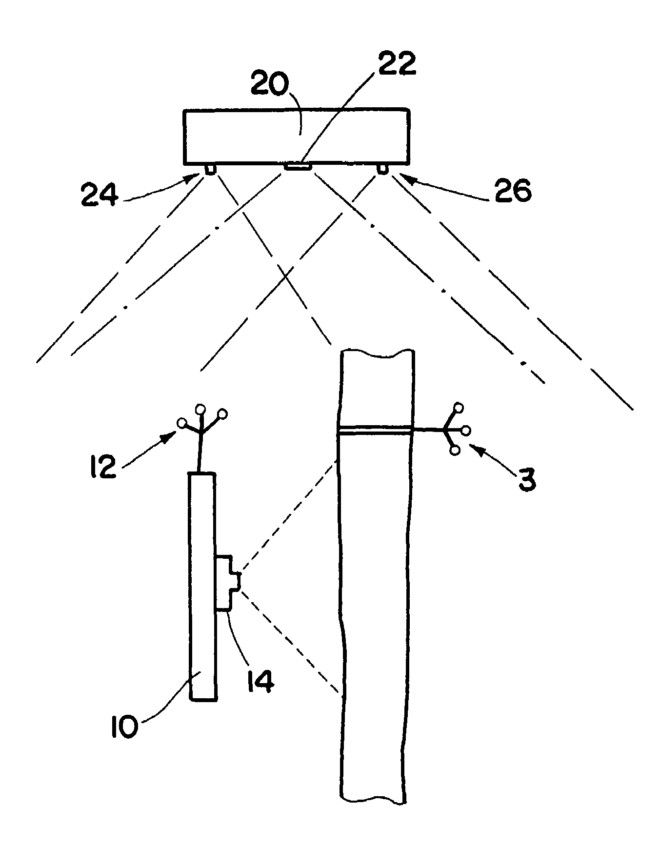
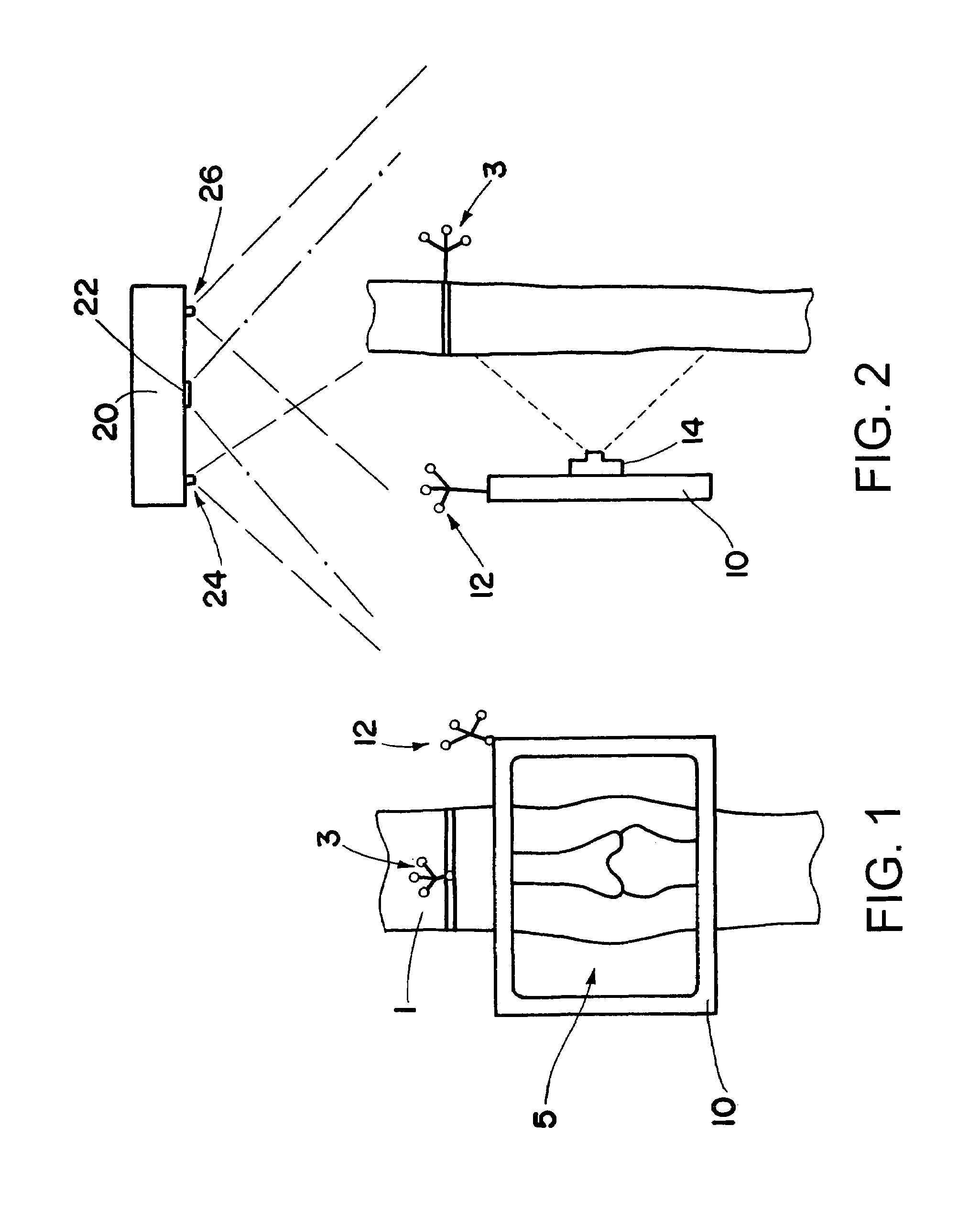
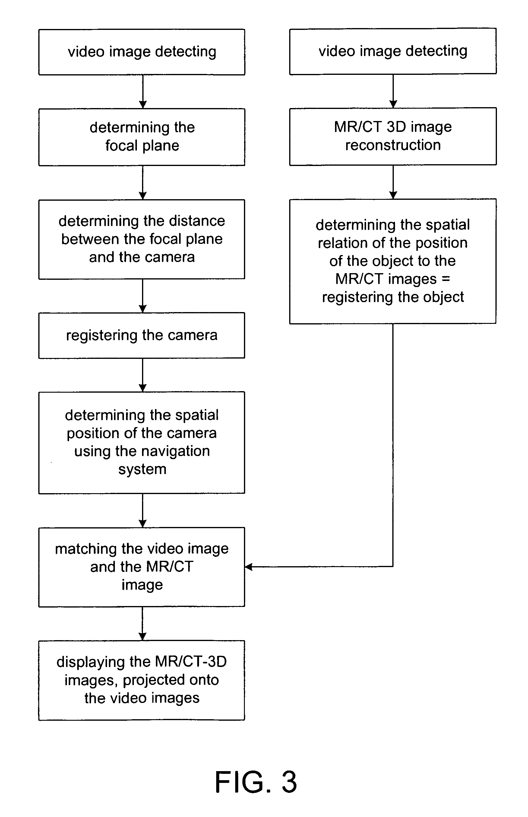
[0018]FIG. 1 schematically shows a device in accordance with the invention, using which the interior of a patient's joint may for example be inspected. The leg 1 of the patient is to be examined at the joint 5. To this end, the flat screen 10 designed in accordance with the invention is held directly in front of the joint, and an image then appears on the screen 10 containing both image information from the video camera image of the camera 14 (FIG. 2) as well as image information on the interior anatomical structure, said information being communicated for example by a navigation apparatus, by radio. To this end, a tomographic image (MR or CT image) is produced beforehand of the patient or of the patient's leg 1.

[0019]As is indicated more clearly in FIG. 2, the camera 14 is situated, firmly attached, on the rear side of the screen 10.

[0020]This type of image representation is made possible by the spatial position of the camera 14 or of the image screen 10 being tracked around the le...


PUM


Abstract

The invention relates to a device for projecting patient image data from radioscopic imaging methods and / or tomographic imaging methods onto video images, comprising an image display device (10), at least one video camera (14) and a computer-assisted navigation system which can detect the spatial positions of the image display device (10) and / or the video camera (14) and the spatial positions of a part of a patient's body (1) via tracking means (12, 3) attached to it, wherein the image display device (10) and the video camera (14) are assigned to each other and are realized as a portable unit.

Description

[0001]The present invention relates to a device for projecting patient image data from radioscopic imaging methods and / or tomographic imaging methods onto video images. The device comprises an image display device, at least one video camera and a computer-assisted navigation system which can detect the spatial position of the image display device and / or the video camera and the spatial positions of a part of a patient's body via tracking means attached to it.[0002]Such devices serve to visually assist a physician during diagnosis or while treating a patient. The option should be provided of combining images from the patient's interior with conventional video images, in order to facilitate treatment and diagnosis.BACKGROUND OF THE INVENTION[0003]A video-based surgical target system is known from U.S. Pat. No. 5,765,561, for which among other things the use of a tracked video camera is proposed, whose images are superimposed onto assigned images from radioscopic imaging methods and / or...

Claims


Application Information

Patent Timeline
IPC IPC(8): A61B5/05A61B5/055A61B6/00A61B34/20A61B90/00A61B90/30
CPCA61B6/00A61B6/461A61B6/462A61B6/547A61B19/52A61B19/5244A61B5/055A61B2019/5291A61B19/5212A61B2019/5229A61B2019/5255A61B2019/5272A61B2019/5289A61B19/5202A61B90/36A61B2034/2055A61B2034/2072A61B34/20A61B90/30A61B2090/364A61B90/361A61B2090/372A61B2090/365
Inventor VILSMEIER, STEFAN
Owner BRAINLAB
Why Eureka
Try Eureka