
Method and apparatus for tissue harmonic imaging with natural (tissue) decoded coded excitation

a harmonic imaging and tissue technology, applied in the field of tissue harmonic imaging using an ultrasound machine, can solve the problems of limiting the bandwidth of the probe, the use of a narrowed transmit fundamental band, and so as to improve the harmonic imaging performance, avoid the frame rate drop, and increase the penetration and snr

Inactive Publication Date: 2006-05-02
GE MEDICAL SYSTEMS INC
View PDF6 Cites 12 Cited by
  • Summary
  • Abstract
  • Description
  • Claims
  • Application Information

AI Technical Summary

Benefits of technology

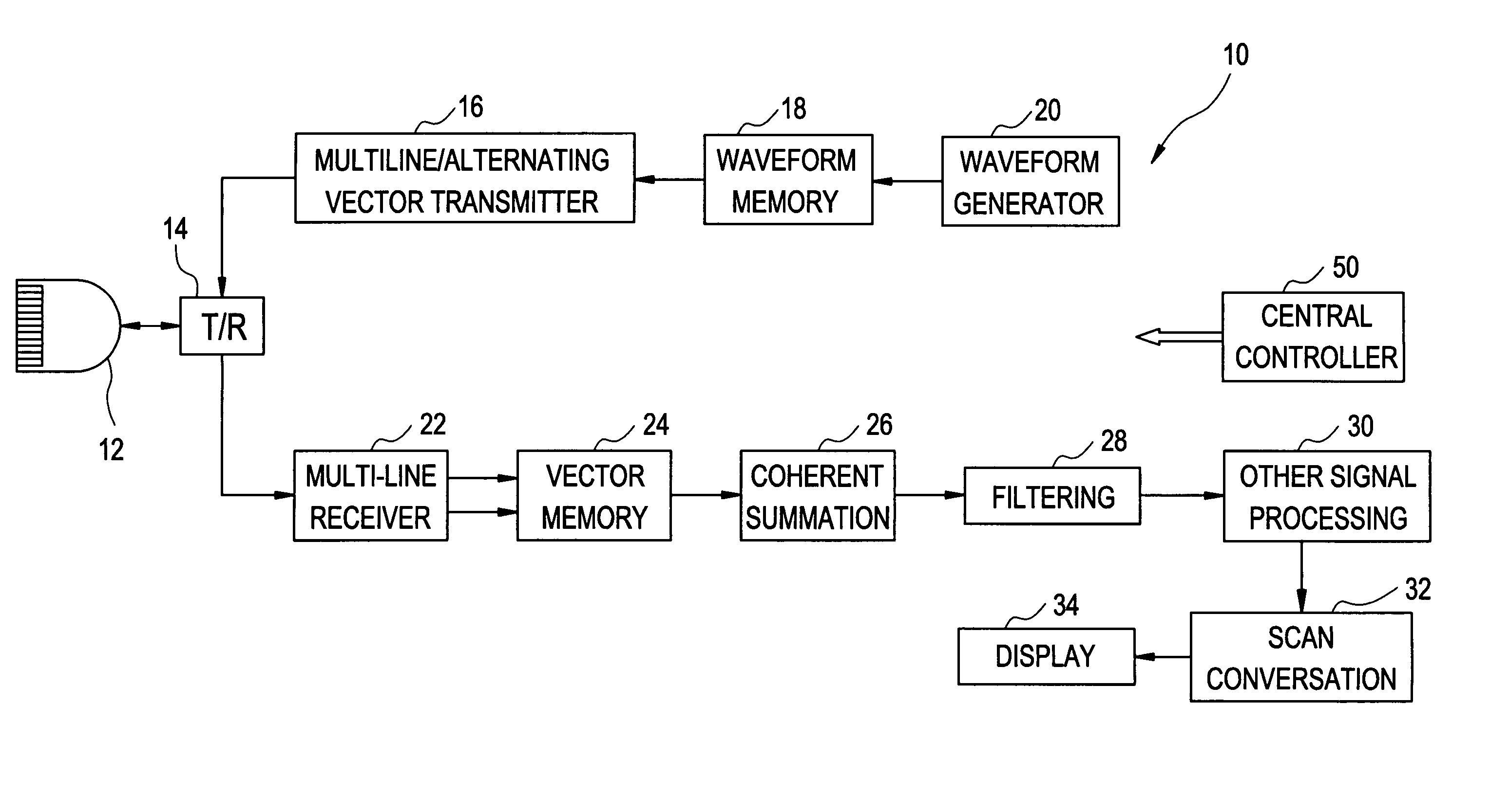
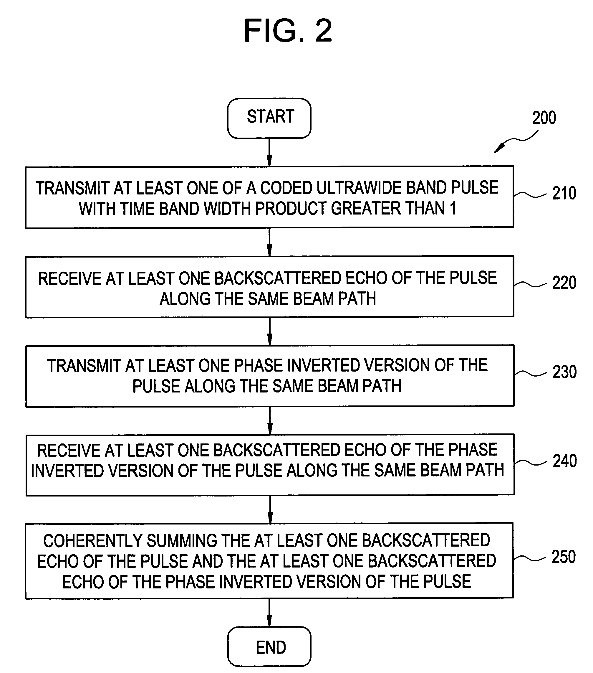
[0012]One embodiment of the present invention relates to tissue harmonic imaging using an ultrasound machine. At least one embodiment of the invention uses wideband (BW greater than about 80%) coded excitation transmit signals with time bandwidth product greater than about 1 for harmonic imaging. At least one embodiment employs pulse inversion for removing fundamental signals, and further employs at least one of the Multi-line acquisition, Phase alternating in space, and Multi-line transmission methods (or a combination of two or more of such methods) to avoid the frame rate drop caused by double firing as provided previously. The wideband transmit signal improves the harmonic imaging performance in very near field while at the same time increases the penetration and SNR compared to a normal pulse inversion method where regular pulses (time bandwidth product=1) are employed. Decoding of the receiving signal is naturally implemented through propagation of coded pulses inside tissue and pulse inversion.
[0013]It is contemplated that embodiments of this invention may greatly improve the image resolution compared to single firing band pass filtering harmonic imaging method while maintaining the same or even better penetration and SNR as in single firing harmonic imaging, which greatly surpass the penetration of regular pulse inversion harmonic imaging.

Problems solved by technology

However, using band pass filtering may limit the bandwidth.
The limitation of the probe bandwidth will force the use of a narrowed transmit fundamental band.
A narrow bandwidth may require lengthening the transmit pulse and filter impulse response, which, in turn, may result in a degradation of axial resolution.
However, for regular pulses, broadband means short pulse length, which in turn, results in a loss of penetration especially in harmonic imaging where the second harmonic signal is about 20 dB less than the fundamental signal.
Further, requirement of double or multiple firing along the same beam path reduce the frame rate in phase inversion case.
However, several major challenges are contemplated.
First, it is always a challenge to obtain sufficient penetration and improved SNR while maintaining good resolution.
In both cases, the decoding filter is quite large and costly.
The cost issue prevents most of the current ultrasound companies from implementing the frequency / nonlinear phase modulated coded excitation even in their premium ultrasound machines.
Even if the cost were acceptable, the decoding filter with a matched design may not work as planned most of the time, especially for harmonic imaging.
This is due to the fact that in harmonic imaging, the decompressing becomes more difficult since the phases change twice as fast, as a function of time, compared to the fundamental, and the complexity of the generation of tissue harmonic through generally heterogonous tissue severely degrade the effectiveness of the matched compression filtering, which is always designed based on ideal situations.
As a result, the range side lobe level could be very high, which does not make sense for practical applications of ultrasound imaging.
Another challenge in harmonic imaging is the near field harmonic performance.
Thus, for very near field (e.g., less than about 2 cm) there may not be enough harmonic components generated in tissue.
Generally, this causes blooming-like image characteristics where a thin line could appear as a thick block due to either saturation of the over-gained small second harmonic signal or leakage of the fundamental signal especially in high frequency case.
This phenomenon seriously limits the application of harmonics in near field structure such as small parts and superficial structure with high frequency probes.
Phase inversion techniques generally sacrifice frame rate since multiple firings are required per beam position.

Method used


Image

  • Method and apparatus for tissue harmonic imaging with natural (tissue) decoded coded excitation
  • Method and apparatus for tissue harmonic imaging with natural (tissue) decoded coded excitation
  • Method and apparatus for tissue harmonic imaging with natural (tissue) decoded coded excitation

Examples


Embodiment Construction

[0033]For the purpose of illustration only, the following detailed description references a certain embodiment of an ultrasound machine, apparatus or device. However, it is understood that the present invention may be used with other devices or imaging systems.

[0034]One or more embodiments of the present invention attempts to solve the three challenges discussed previously with respect to current tissue harmonic imaging implementations: 1) trade-off between penetration, SNR and resolution; 2) near field harmonic performance; and 3) frame rate drop with multiple firing for higher resolution. Embodiments of the present invention solve these challenges using frequency modulated coded excitation pulses in combination with pulse inversion, where the waveforms have a time bandwidth product greater than about 1, and a bandwidth greater than about 80%. It is contemplated that no costly matched decoding / compressing filter are used for decoding.

[0035]FIG. 1 illustrates an ultrasound apparatus...


PUM


Abstract

The present invention relates to a method and apparatus providing tissue harmonic imaging using an ultrasound machine. Coded pulses and the phase inverted version of the said coded pulses with time bandwidth greater than 1 are transmitted into the tissue. Backscattered echoes are received and filtered before or after coherent summation. Decoding / compressing of the received echoes of the coded pulses is implemented naturally through the propagation of the specially designed ultrawide band (>80%) waveforms inside tissue and pulse inversion. Costly decoding / compression filter are not necessary.

Description

RELATED APPLICATIONS[0001]This application is related to, and claims benefit of and priority from, Provisional Application No. 60 / 501,784 filed on Sep. 9, 2003, titled “Method and Apparatus for Tissue Harmonic Imaging with Natural (Tissue) Decoded Coded Excitation”, the complete subject matter of which is incorporated herein by reference in its entirety.FEDERALLY SPONSORED RESEARCH OR DEVELOPMENT[0002][Not Applicable]MICROFICHE / COPYRIGHT REFERENCE[0003][Not Applicable]BACKGROUND OF THE INVENTION[0004]This application is directed in general to imaging using an ultrasound machine. This application is directed in particular to tissue harmonic imaging using an ultrasound machine.[0005]Tissue harmonic imaging is a known imaging method used in ultrasound machines. Such imaging was started based on a perceived need to improve the effectiveness of acoustic contrast agents, where the backscattered signal is rich in harmonics. Once introduced into clinical practice, it became obvious that ima...

Claims


Application Information

Patent Timeline
Patent Type & Authority Patents(United States)
IPC IPC(8): A61B8/00G01S7/52G01S15/89
CPCG01S7/52038G01S7/52093G01S15/8963G01S15/8959G01S7/52095
Inventor HAO, XIAOHUICHIAO, RICHARDMILLER, STEVENLI, YADONG
Owner GE MEDICAL SYSTEMS INC
Learn More