
Flexible endoscope capsule

a flexible, endoscope technology, applied in the field of minimally invasive internal surgery, can solve the problems of difficult and sometimes tedious task of surgical personnel to complete medical procedures in endoscopic applications, the tubular members utilized in endoscopic applications are necessarily of flexible construction and may be of significant length, and achieve the effect of facilitating the performance of endoscopic medical procedures

Inactive Publication Date: 2005-06-21
PARE SURGICAL
View PDF9 Cites 307 Cited by
  • Summary
  • Abstract
  • Description
  • Claims
  • Application Information

AI Technical Summary

Benefits of technology

[0005]An objective of the present invention is to provide an apparatus and method that facilitates the performance of endoscopic medical procedures.
[0006]Another objective is to provide an apparatus and method for facilitating the performance of endoscopic medical procedures in a minimally complex manner.
[0007]A further objective is to provide an apparatus and method that facilitates performing endoscopic medical procedures via an approach that is compatible with existing endoscopic devices.

Problems solved by technology

Given such access points and associated bodily canals, the tubular members utilized in endoscopic applications are necessarily of flexible construction and may be of significant length.
To date, endoscopic procedures have been largely limited to gasdroesophergeal (GERD) and biopsy applications.
As may be appreciated, the completion of medical procedures in endoscopic applications can present a challenging and sometimes tedious task for surgical personnel.
For example, suturing procedures may involve difficult manipulation of an external device to cause an internally located needle to pass entirely through tissue at a surgical site to effect suture stitching.
An additional problem that may be encountered during these endoscopic medical procedures is that while the endoscope is inserted within the hollow cavity of an organ, these organs are generally in a flaccid or relaxed state.
In this regard, a physician may have a limited field of view of a tissue site area of interest as well as little room in which to manipulate medical instruments relative to the tissue site to perform a desired medical procedure.
In addition, relaxed tissue does not provide a structural surface against which the endoscope may apply pressure during a procedure.

Method used


Image

  • Flexible endoscope capsule
  • Flexible endoscope capsule
  • Flexible endoscope capsule

Examples


first embodiment

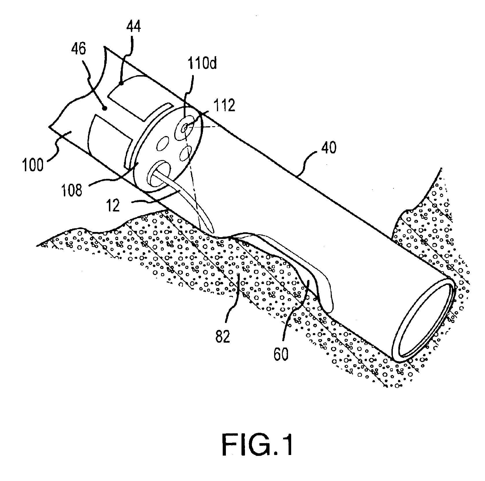
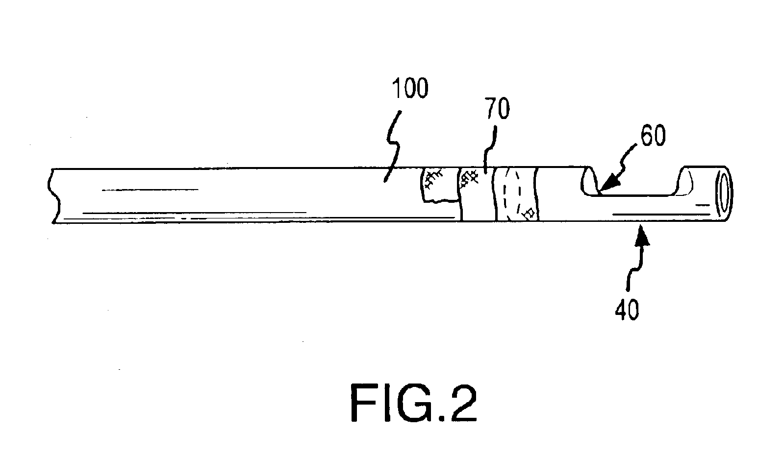
[0037]FIGS. 1 and 2 illustrate an end cap 40 that may be interconnected to an endoscopic device 100. The end cap 40 provides an improved field of view for endoscopic imaging device 112 as well as working space for endoscopic medical instruments to perform medical procedures. As used herein, the term endoscopic device includes a plurality of minimally invasive surgical devices (i.e., scopes) that have been developed for specific uses. For example, upper and lower endoscopes are utilized for accessing the esophagus / stomach and the colon, respectively, angioscopes are utilized for examining blood vessels, and laparoscopes are utilized for examining the peritoneal cavity. Though discussed herein in relation to use with endoscopic devices such as the type utilized for colon and esophageal applications, it will be appreciated that numerous other embodiments including one or more aspects of the present invention may be constructed for use with any minimally invasive surgical device.

[0038]T...

second embodiment

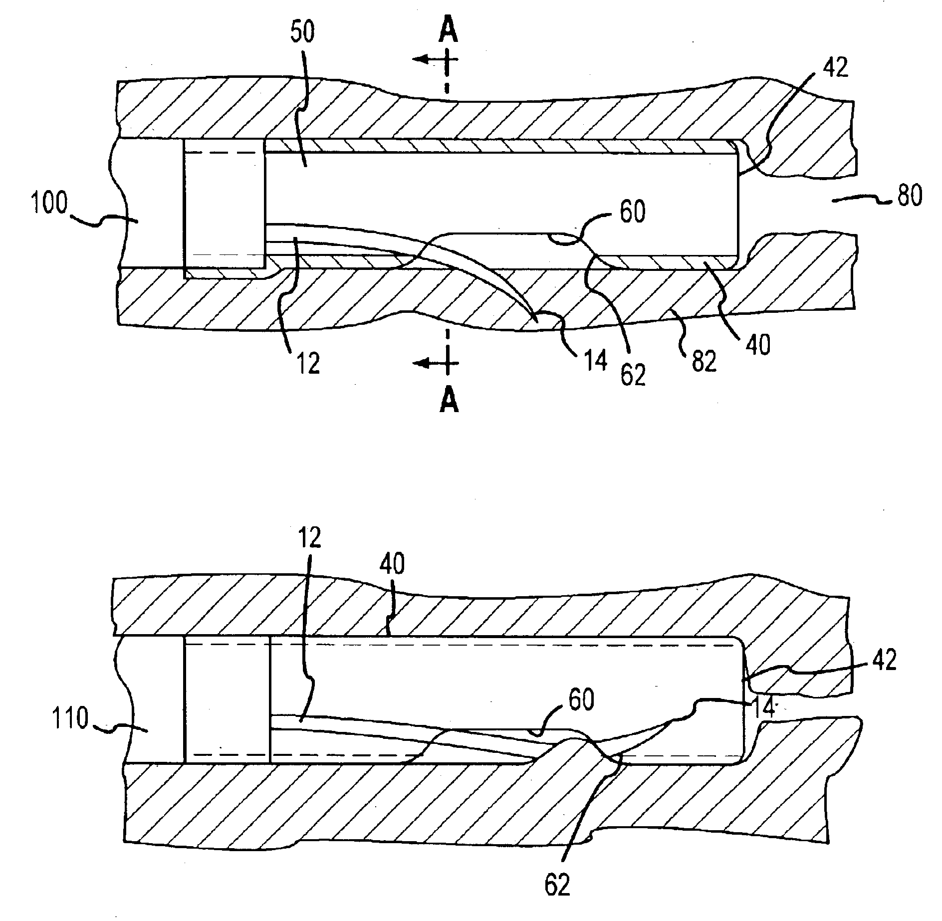
[0042]As noted, the attachment end 44 of the substantially hollow cylindrical end cap 40 is open defining an aperture to receive the distal end 108 of an endoscopic device 100. In this regard, the end cap 40 defines an internal chamber 50 that is accessible by endoscopic instruments through the distal end 108 of the endoscopic device 100. Referring to FIG. 3a, a cross-sectional view of the end cap 40 interconnected to the distal end of the endoscopic device 100 is shown. As illustrated, the attachment end 44 of the end cap 40 receives the distal end of the endoscopic device 100. Accordingly, medical instruments, such as a needle 12, can access the internal chamber 50 of the end cap 40. In the embodiment of FIGS. 3a and 3b, the distal end 42 of the end cap 40 is open. This allows suction or irrigation provided by the endoscopic device 100 to pass through the end cap 40 unimpeded. FIGS. 4a and 4b show the end cap 40 where the distal end 42 of the end cap 40 is closed to allow suction ...


PUM


Abstract

An endoscopic capsule adapted for selective attachment to the distal end portion of an endoscopic device. The capsule is designed for insertion with an interconnected endoscopic device into a patient body to provide an open space in front of the distal end of the endoscopic device. The capsule facilitates the performance of endoscopic procedures providing a space in which to manipulate medical instruments within the patient body as well as improving a surgeon's field of vision within the patient body. In one embodiment, the capsule includes a housing having an internal chamber and two apertures. A first aperture is interconnectable to the end of an endoscopic device and provides operative access to endoscopic instruments contained therein. One or more of these endoscopic devices are advanceable into the internal cavity for selectively engaging patient tissue disposed relative to the second aperture.

Description

FIELD OF THE INVENTION[0001]The present invention relates to the field of minimally invasive internal surgery, and more particularly, to facilitating endoscopic procedures.BACKGROUND OF THE INVENTION[0002]Minimally invasive internal surgery procedures are ever-increasing. Such procedures typically entail the insertion of one or a plurality of tubular members into a patient body and the passage of various devices through the tubular member(s) to access a tissue site of interest.[0003]In certain endoscopic procedures, a tubular member is inserted through a bodily orifice to provide instrumentation access therethrough to an internal tissue site, e.g., through the mouth or anus to access the “lumen” or cavity of a hollow organ. The tubular member generally contains a plurality of individual channels extending along its entire length for providing, inter alia, suction, water, and / or air as well as providing access for optical equipment and medical instruments. Given such access points an...

Claims


Application Information

Patent Timeline
IPC IPC(8): A61B1/012A61B1/018
CPCA61B1/00087A61B1/018A61B2017/00269
Inventor FLEENER, RICHARD P.BROMLEY, ROBERT L.
Owner PARE SURGICAL
Learn More