Method and system for automatic classification and quantitative evaluation of adnexal masses based on a cross-sectional or projectional images of the adnex
- Summary
- Abstract
- Description
- Claims
- Application Information
AI Technical Summary
Problems solved by technology
Method used
Image


Examples
example 1
Defining Mass Boundaries
As mentioned hereinabove, numerous methods in image analysis pan be employed to define the boundaries of an image component which differs from its surroundings. This example describes a prior art process [15] which was successfully applied to define boundaries of ovarian cysts.
Segmentation is often an important step in the analysis of medical images. The gray value of a pixel is the most widely used variable for region segmentation. Other features, such as the local texture, can also be utilized for that purpose. The local entropy, however, is considered more suitable for boundary extraction. Nevertheless, in ultrasound images containing transparent fluid encircled by soft tissue it can be used for region segmentation. Although much progress has been accomplished in automatic segmentation of medical ultrasound images, gynecological images received relatively less attention. As an example, automatic segmentation applied on the ovaries can only be rarely found ...
example 2
Computerized Quantification of Structures within Ovarian Cysts Using Ultrasound Images
Ovarian cysts are a common type of ovarian mass. The morphology of cysts, as it appears in ultrasound images, is currently used for classification of ovarian pathologies. However, this classification process is based on human interpretation of the sonographic image. In this Example, a semi-automatic algorithm for the quantification of ovarian cysts is presented. This algorithm categorizes the structures within a cyst and extracts their quantitative geometrical properties (e.g., width, characteristic diameter). In order to assess the validity of the technique, its performance was compared to human classification and manual measurements made by an expert. The results show a good match between automatic evaluations made by a computer and those of an experienced observer, indicating a potential for clinical use.
As already mentioned, ovarian cysts are the most common type of ovarian mass observed in the...
example 3
The Convex Hull
The convex hull of a shape is the smallest convex object that contains the original shape. The area added to the original shape (i.e., the difference between the original object and its convex hull) is called the convex deficiency. The convex hull is extensively discussed in the literature, and numerous algorithms have been proposed in order to compute it. A variety of algorithms for finding the convex hull of a finite set of points in the plan is summarized by Preparata and Shamos [31] and Day [32]. Although a shape in a binary image can be viewed as a finite set of points (located at the centers of the pixels), relatively few techniques addressed the convex hull in images.
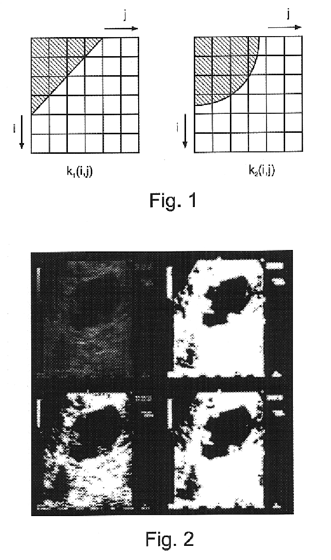
The upper convex hull (U-hull) of a digital planar object is composed of straight segments bounding the object from above. Similarly, its lower convex hull (L-hull) is composed of straight segments bounding it from beneath. The complete convex hull of the shape can be obtained by combining the U-hu...
PUM
 Abstract
Description
Claims
Application Information
 - R&D
- Intellectual Property
- Life Sciences
- Materials
- Tech Scout
- Unparalleled Data Quality
- Higher Quality Content
- 60% Fewer Hallucinations
Browse by: Latest US Patents, China's latest patents, Technical Efficacy Thesaurus, Application Domain, Technology Topic, Popular Technical Reports.
© 2025 PatSnap. All rights reserved.Legal|Privacy policy|Modern Slavery Act Transparency Statement|Sitemap|About US| Contact US: help@patsnap.com