Apparatus and method for compressing body tissue

Inactive Publication Date: 2005-02-01
OVESCO ENDOSCOPY AG
View PDF3 Cites 430 Cited by
  • Summary
  • Abstract
  • Description
  • Claims
  • Application Information

AI Technical Summary

Problems solved by technology

However, surgery is not preferred unless there is no endoscopic alternative or if previous endoscopic efforts have not succeeded.
Surgical intervention is not preferred for at least the reasons that it has associated with it greater morbidity and mortality, and also, significantly higher costs than other procedures.
Untreated visible vessels are likely to bleed.
For the GI endoscopist, hemorrhaging is the most worrisome procedure.
It is the one problem the endoscopist faces that is generally not an outpatient procedure.
These rates have not improved significantly in decades.
However, drawbacks exist with these known procedures for the endoscopist.
However, both methods require a specialized power generator.
For mechanical compression, loops and mechanical clips are known for use, however, problems exist with each.
Whereas the loop is an endoscopically delivered compression devices its primary use is for bleeding polyp stalks, and thus, it is not designed for, nor appropriate for use in, ulcer treatment procedures.
A mechanical clip is known, however, the known mechanical clip has drawbacks.
Additionally, the clip is not able to provide sufficient clamping force because of its structural design.
Thus, these clips require multiple applications and are not effective for definitive hemostasis.
An additional problem with these clips is that when delivering these clips to the wound site, good visualization of the bleeding vessel cannot be obtained.
The endoscopist may be required to blindly attach the clip, resulting in an imprecisely preformed procedure that may require guess work on the part of the endoscopist.

Method used


Image

  • Apparatus and method for compressing body tissue
  • Apparatus and method for compressing body tissue
  • Apparatus and method for compressing body tissue

Examples


first embodiment

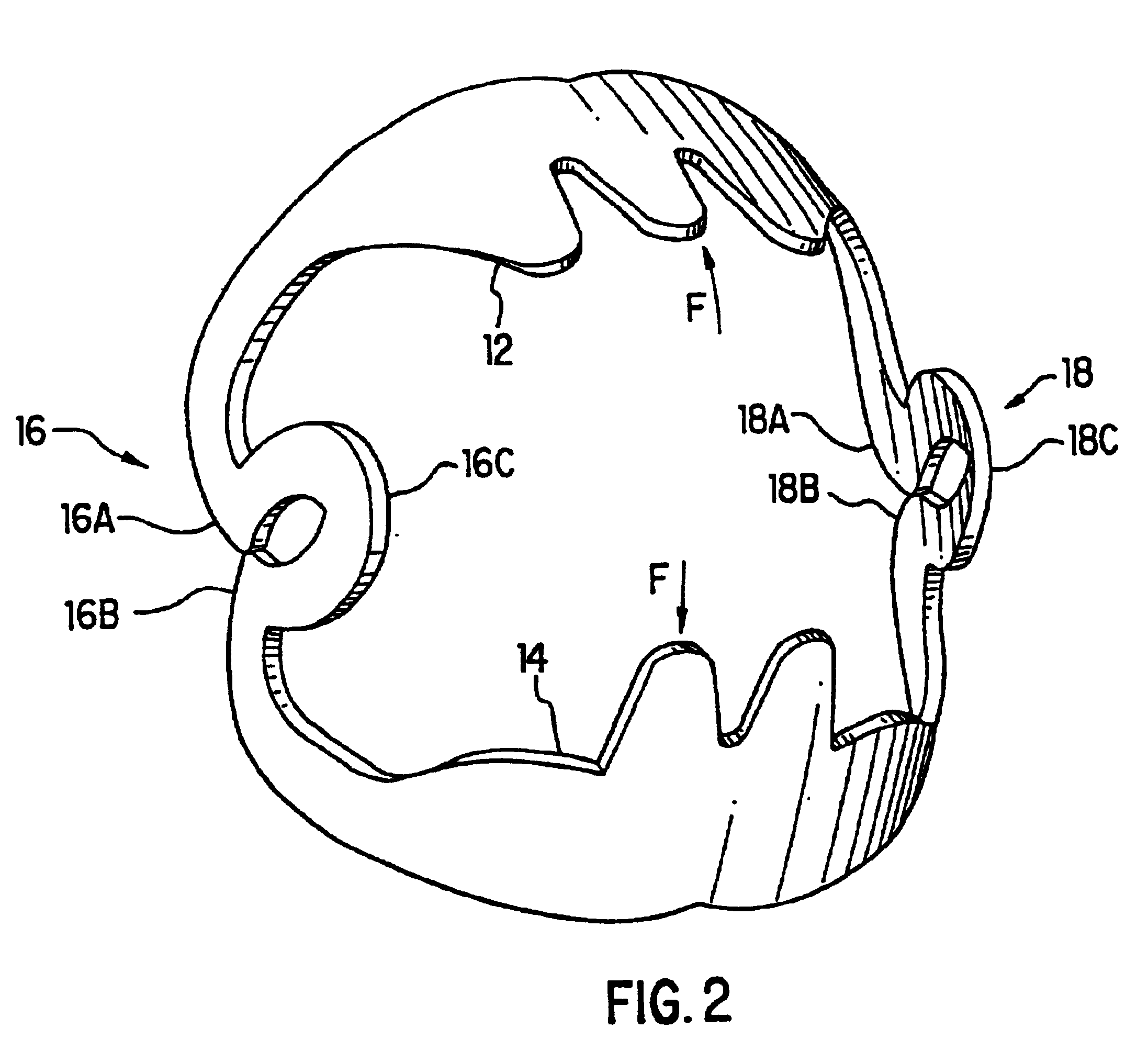
FIG. 1 illustrates a first embodiment for a surgical clip that may be delivered to a site within a patient's body by an endoscopic device. The system and method for delivering surgical clip 10 to the wound site in the patient's body will be discussed later in this specification.

As can be seen in FIG. 1, surgical clip 10 is comprised of a first elongated tissue grasping surface 12 which has a first end 12A and a second end 12B and a second elongated tissue grasping surface 14 also having a first end 14A and a second end 14B. As can also be seen in FIG. 1, both the first tissue grasping surface 12 and the second tissue grasping surface 14 are formed by a semi-circular member.

A first joint 16 and a second joint 18 connect first elongated tissue grasping surface 12 to second elongated tissue grasping surface 14. First joint 16 is connected at a first end 16A to the first end 12A of first elongated tissue grasping surface 12 and at a second end 16B to the first end 14A of second tissue g...

third embodiment

FIG. 6 illustrates a third embodiment for a deployment device. In the embodiment of FIG. 6, the deployment device comprises a balloon 300 where at least a portion of balloon 300 is disposed between surgical clip 10 and endoscope cap 4. An inflation lumen 310 extends from balloon 300 to a position outside of the patient such that pressure may be applied to balloon 300 to inflate balloon 300. Any substance may be utilized to inflate the balloon, including a gas, liquid, or any other substance. As can be understood, balloon 300 may be maintained in a state of inflation such that balloon 300 does not force surgical clip 10 off of endoscope cap 4. When the surgeon desires to deploy surgical clip 10 from endoscope cap 4, the surgeon would inflate balloon 300 to a state such that the inflation of balloon 300 causes surgical clip 10 to be moved toward the distal end of endoscope cap 4 such that continued inflation of balloon 300 will deploy surgical clip 10 off of endoscope cap 4.

As the bal...

fourth embodiment

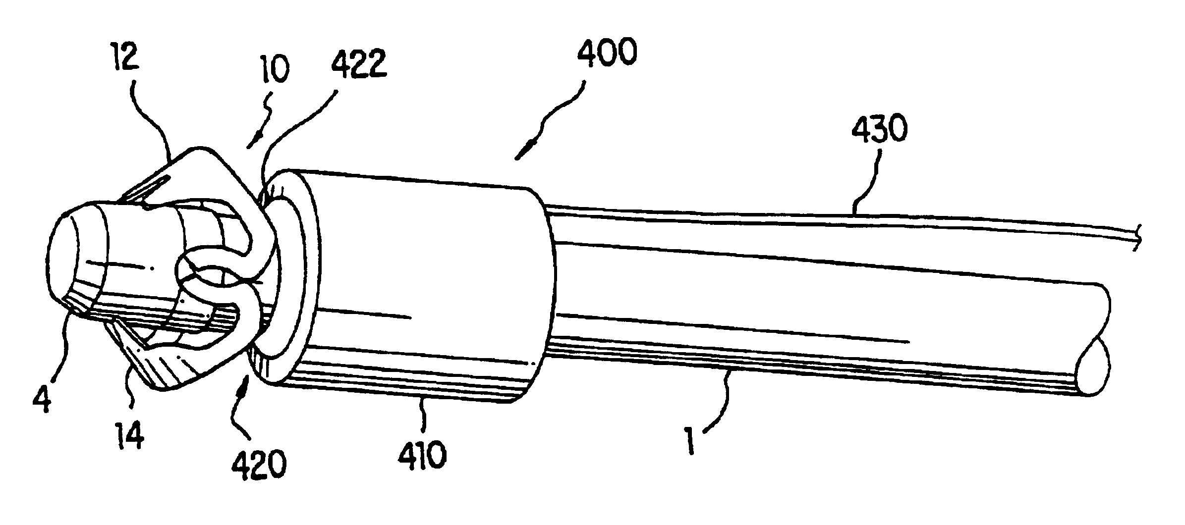
FIG. 7 illustrates a deployment device 400 that incorporates a force generator 410 that is disposed around endoscopic device 1 and is located proximal to surgical clip 10. As can be seen in FIGS. 7 and 8, force generator 410 includes an engagement member 420 that is at least partially disposed within the force generator 410 and which is movable between a first position where a distal end 422 of engagement member 420 does not engage with surgical clip 10 and a second position where distal end 422 of engagement member 420 engages with surgical clip 10. An actuator 440 is contained within force generator 410 for moving engagement member 420 to its second position where it engages with surgical clip 10.

FIG. 8 is a cross sectional view that further illustrates the fourth embodiment for deployment device 400 that includes force generator 410. As can be seen in FIG. 8, engagement member 420 is at least partially disposed within force generator 410. A retention spring 425 may be utilized to...


PUM


Abstract

A system and method for delivering a surgical clip to a surgical site within a patient's body to compress body tissue is disclosed. In one embodiment for the system of the present invention, the system includes an endoscopic device that has an endoscope cap disposed on a distal end of the endoscopic device. A surgical clip is removably disposed on an outside surface of the endoscope cap. A deployment device is associated with the surgical clip for deploying the surgical clip from the endoscope cap to the body tissue that is to be compressed.

Description

BACKGROUND OF THE INVENTION1. Field of the InventionThe present invention relates generally to an apparatus and method for compressing body tissue to prevent hemorrhaging at a surgical site within a patient's body. More specifically, the invention provides a clip and a system for delivering the clip to the surgical site. The present invention could be utilized for any of a variety of procedures, including to close an organ perforation from inside a lumen by approximating and compressing the wound edges of the perforated tissue.2. Description of the Related ArtBleeding Peptic Ulcer Disease can be a critical event since there is internal hemorrhaging associated with the ulcer. Patients that are suspected of having bleeding peptic ulcer disease can be diagnosed and treated endoscopically in emergency rooms of medical centers, intensive care units, or, in a Gastro-Intestinal (GI) suite, although personnel and equipment may need to be transported to the patient. Surgery, either laparosco...

Claims


Application Information

Patent Timeline
IPC IPC(8): A61B17/122A61B17/12A61B17/128A61B17/03A61B17/00A61B17/08
CPCA61B17/10A61B17/122A61B17/1285A61B17/1227A61B17/0057A61B17/083A61B2017/12004A61B2017/00539A61B2017/00544A61B2017/00557
Inventor DURGIN, RUSSELL F.SCHURR, MARC O.BUESS, GERHARDVOGEL, BERNDFISCHER, HARALD
Owner OVESCO ENDOSCOPY AG
Why Eureka
Try Eureka