Immunologic test method and immunologic test kit
a technology of immunologic test and immunological test, which is applied in the direction of instruments, biochemistry apparatus and processes, material analysis, etc., can solve the problems of lack of rapidness and insufficient detection sensitivity
- Summary
- Abstract
- Description
- Claims
- Application Information
AI Technical Summary
Benefits of technology
Problems solved by technology
Method used
Image


Examples
example a-2
Detection of Escherichia coli O157:H7 by Kit for Immunological Detection
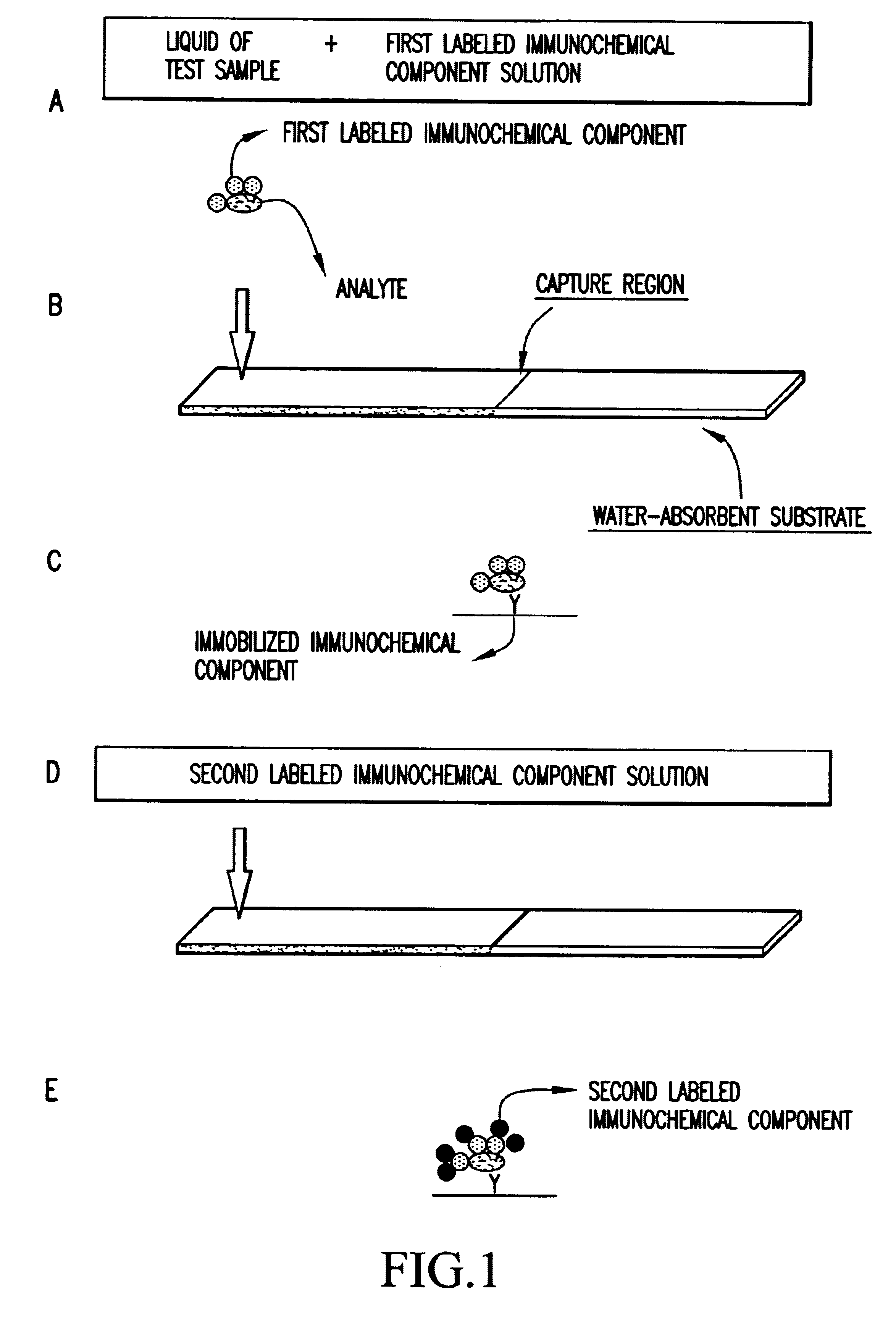
There was prepared a liquid of a test sample obtained from dispersion of Escherichia coli O157:H7 strain in 0.1 M phosphate buffer (pH 7.4) containing 0.9% by weight NaCl at each concentration shown in Table A-1. The resulting liquid of a test sample was mixed with the first labeled antibody solution prepared in item (1) of Example A-1 so as to have a concentration of 0.02% by weight on a solid basis, and the mixture was stirred. Thereafter, 60 .mu.l of a mixture was added dropwise to a polyester nonwoven fabric portion of the test strip prepared in item (3) of Example A-1. The mixture was developed on the test strip, and thereafter 60 .mu.l of a diluted solution was added dropwise to the above polyester nonwoven fabric portion, the diluted solution being prepared by diluting the second labeled antibody solution prepared in item (2) of Example A-1 with 0.1 M phosphate buffer (pH 7.4) containing 0.9% by weight Na...
example a-3
Detection of Escherichia coli O157:H7 by Kit for Immunological Detection
There was prepared a liquid of a test sample obtained from dispersion of Escherichia coli O157:H7 strain in 0.1 M phosphate buffer (pH 7.4) containing 0.9% by weight NaCl at each concentration shown in Table A-1. The resulting liquid of a test sample was mixed with the first labeled antibody solution prepared in item (1) of Example A-1 and the second labeled antibody solution prepared in item (2) of Example A-1 so as to have a concentration of 0.02% by weight each on a solid basis, and the mixture was stirred. Thereafter, 60 .mu.l of the mixture was added dropwise to a polyester nonwoven fabric portion of the immunological test strip prepared in item (3) of Example A-1. The presence or absence of coloring on a capture region after 20 minutes was visually observed. The results are shown in Table A-1. As a comparison, the assay results of the case where only the first labeled antibody was used without using a seco...
example a-4
Detection of Escherichia coli O157:H7 by Kit for Immunological Detection
There was prepared a test sample obtained from dispersion of Escherichia coli O157:H7 strain in 0.1 M phosphate buffer (pH 7.4) containing 0.9% by weight NaCl at each concentration shown in Table A-1. Two microliters of this test sample was allowed to absorb on the front side of the immunological test strip prepared in item (3) of Example A-1 at a site 12 to 20 mm from the opposite side to the antibody-applied site. Subsequently, the first labeled antibody solution prepared in item (1) of Example A-1 was diluted with 0.1 M phosphate buffer (pH 7.4) containing 0.9% by weight NaCl so as to have a concentration of 0.02% by weight on a solid basis. Thereafter, 60 .mu.l of the dilution was added dropwise to a polyester nonwoven fabric portion of the test strip. The first labeled antibody solution was brought into contact with the test sample, and then developed. Thereafter, 60 .mu.l of a diluted solution was added dr...
PUM
| Property | Measurement | Unit |
|---|---|---|
| width | aaaaa | aaaaa |
| width | aaaaa | aaaaa |
| particle size | aaaaa | aaaaa |
Abstract
Description
Claims
Application Information
 - Generate Ideas
- Intellectual Property
- Life Sciences
- Materials
- Tech Scout
- Unparalleled Data Quality
- Higher Quality Content
- 60% Fewer Hallucinations
Browse by: Latest US Patents, China's latest patents, Technical Efficacy Thesaurus, Application Domain, Technology Topic, Popular Technical Reports.
© 2025 PatSnap. All rights reserved.Legal|Privacy policy|Modern Slavery Act Transparency Statement|Sitemap|About US| Contact US: help@patsnap.com