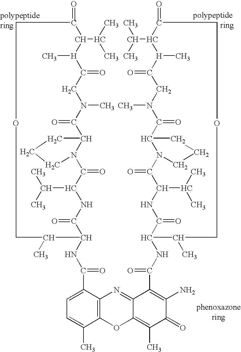
Actinomycin D for the treatment of vascular disease
a technology of vascular disease and actinomycin, which is applied in the direction of blood vessels, prostheses, catheters, etc., can solve the problems of significant morbidity and mortality, recurrent stenosis or restenosis, and remains a significant problem
- Summary
- Abstract
- Description
- Claims
- Application Information
AI Technical Summary
Benefits of technology
Problems solved by technology
Method used
Image
Examples
example 1
Inhibition of SMC Proliferation With Actinomycin D
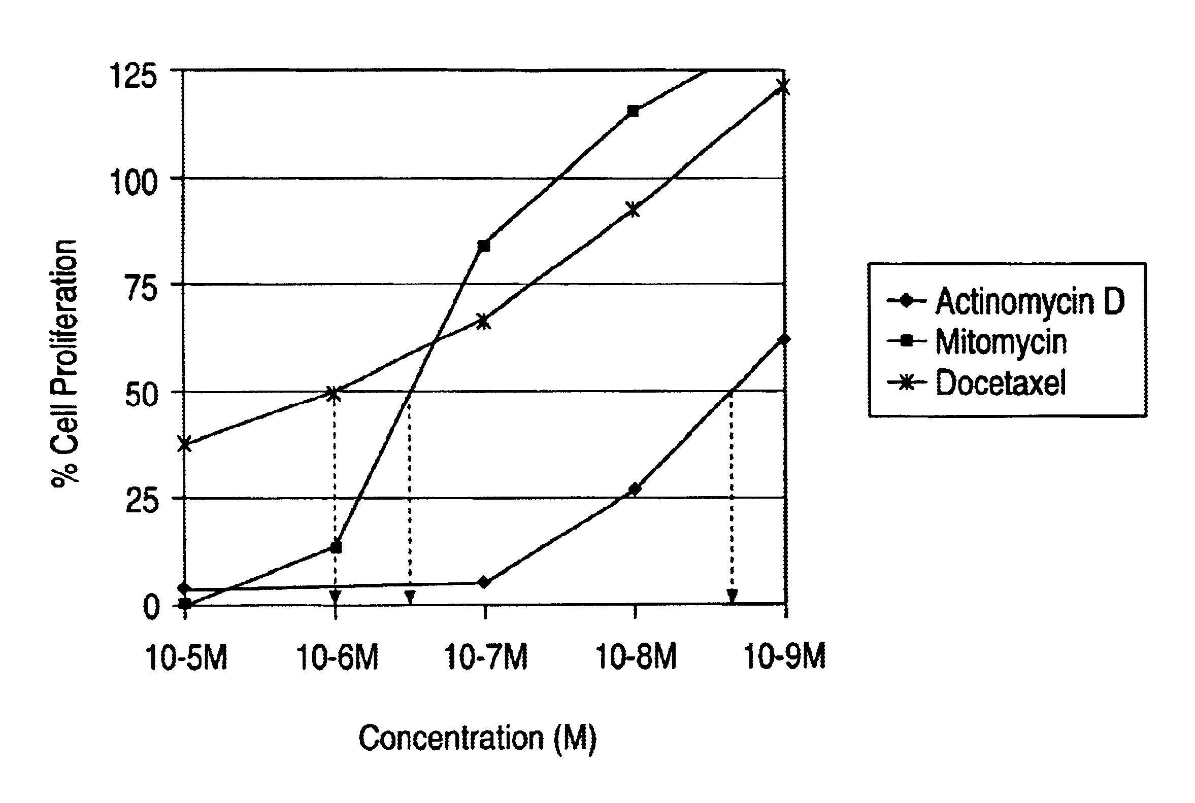
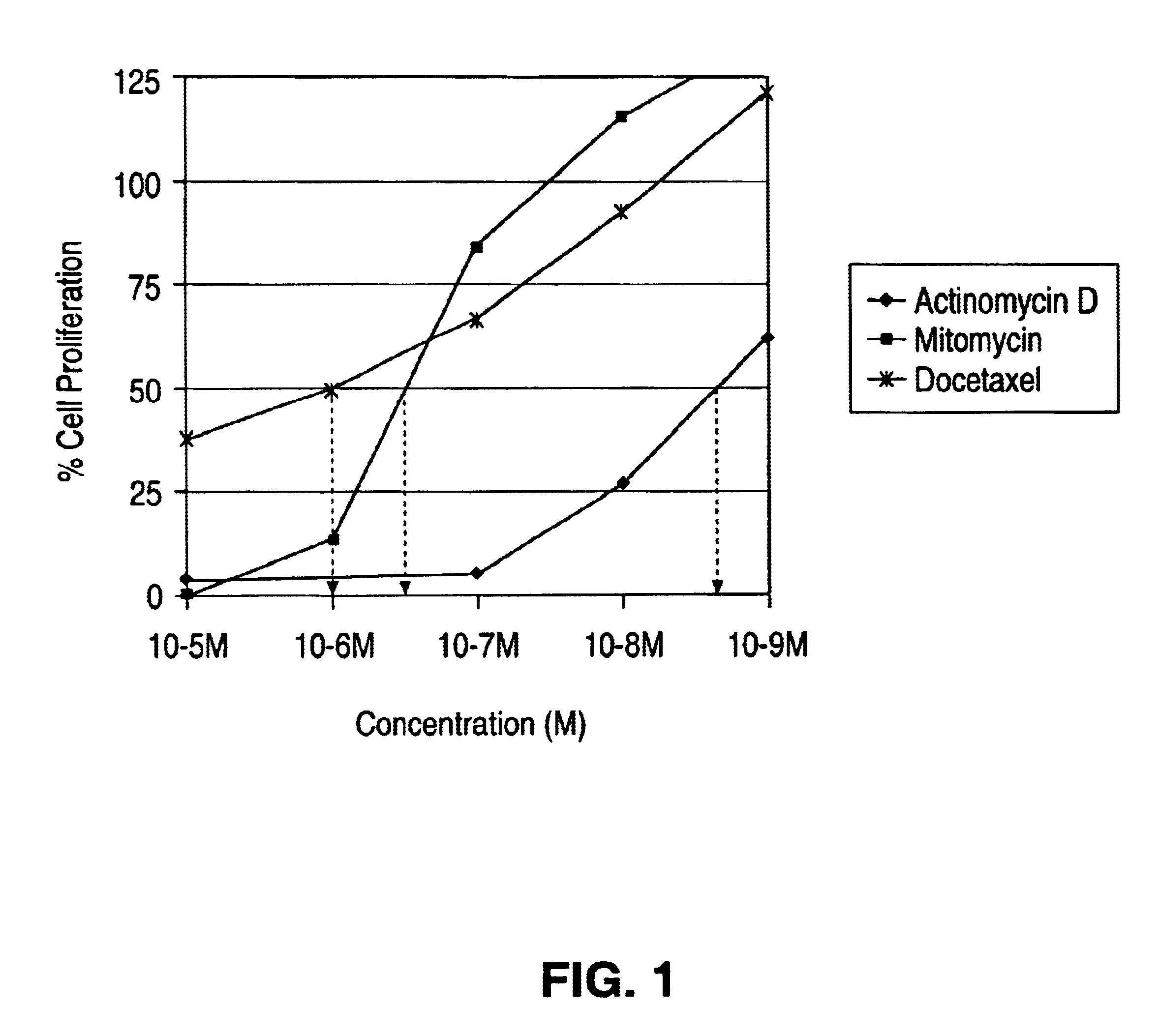
Medial smooth muscle cells (SMC) were isolated from rat aorta and cultured according to explant methods known to one of ordinary skill in the art. Cells were harvested via trypsinization and subcultivated. Cells were identified as vascular SMC through their characteristic hill-and-valley growth pattern as well as indirect immunofluorescence with monoclonal anti SMC .alpha.-actin. Studies were performed with cells at passage 3-4. SMC monlayers were established on 24 well culture dishes, scrape wounded and treated with actinomycin D, mytomycin and docetaxel. The cells were exposed to the drug solution of different concentrations for 2 hours and then washed with buffered saline solution. The proliferation of the cells was quantified by standard technique of thymidine incorporation. The results from the study are tabulated in FIG. 1.
The IC.sub.50 (concentration at which 50% of the cells stop proliferating) of actimomycin D was 10.sup.-9 ...
example 2
Reduction in Restenosis in the Porcine Coronary Artery Model
Porcine coronary models were used to assess the degree of the inhibition of neointimal formation in the coronary arteries of a porcine stent injury model by Actinomycin D, delivered with a microporous balloon catheter (1.times.10.sup.6 pores / cm.sup.2 with size ranging from 0.2-0.8 micron).
The preclinical animal testing was performed in accordance with the NIH Guide for Care and Use of Laboratory Animals. Domestic swine were utilized to evaluate effect of the drug on the inhibition of the neointimal formation. Each testing procedure, excluding the angiographic analysis at the follow-up endpoints, was conducted using sterile techniques. During the study procedure, the activated clotting time (ACT) was monitored regularly to ensure appropriate anticoagulation. Base line blood samples were collected for each animal before initiation of the procedure. Quantitative coronary angiographic analysis (QCA) and intravascular ultrasound...
example 3
In vivo data is provided illustrated positive remodeling caused by the application of actinomycin D. Stents coated with EVAL impregnated with actinomycin D and a control group of stents coated with EVAL free from actinomycin D were implanted in porcine coronary arteries. The animals were sacrificed at the end of 28 days. The EEL area of the actinomycin D-loaded vessels was statistically significantly greater than the EEL area of the control vessels. The index of remodeling was 1.076 (8.54 / 7.94).
Condition Mean Area Std Dev IEL Drug coated(Act-D in EVAL) 7.47 0.89 Control (EVAL) 6.6 0.61 p value 0.0002 Statistical significant difference EEL (external elastic lamia) Drug coated(Act-D in EVAL) 8.54 0.87 Control (EVAL) 7.94 0.73 p value 0.014 Statistical significant difference
EEL Area (mm.sup.2) ID # Control ID # Actinomycin D ID # EVAL 48 LCX d 6.3966 63 LCX d 7.4498 63 LAD d 8.3037 48 LCX m 7.4601 63 LCX m 8.2509 63 LAD m 8.8545 48 LCX p 7.3063 63 LCX p 7.7342 63 LAD p 9.4698 49 LAD d ...
PUM
| Property | Measurement | Unit |
|---|---|---|
| Volume | aaaaa | aaaaa |
| Volume | aaaaa | aaaaa |
| Volume | aaaaa | aaaaa |
Abstract
Description
Claims
Application Information
 - R&D
- Intellectual Property
- Life Sciences
- Materials
- Tech Scout
- Unparalleled Data Quality
- Higher Quality Content
- 60% Fewer Hallucinations
Browse by: Latest US Patents, China's latest patents, Technical Efficacy Thesaurus, Application Domain, Technology Topic, Popular Technical Reports.
© 2025 PatSnap. All rights reserved.Legal|Privacy policy|Modern Slavery Act Transparency Statement|Sitemap|About US| Contact US: help@patsnap.com


