Three-dimensional ultrasound tomography method and system based on spiral scanning
a three-dimensional ultrasound and spiral scanning technology, applied in tomography, ultrasonic/sonic/infrasonic image/data processing, instruments, etc., can solve the problems of low resolution between image layers, difficulty in subsequent three-dimensional reconstruction,
- Summary
- Abstract
- Description
- Claims
- Application Information
AI Technical Summary
Benefits of technology
Problems solved by technology
Method used
Image

Examples
embodiment 1
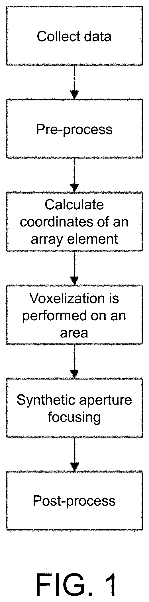
[0050]The three-dimensional ultrasound tomography method based on spiral scanning in the embodiment specifically includes the following steps.
[0051](1) Collecting data:
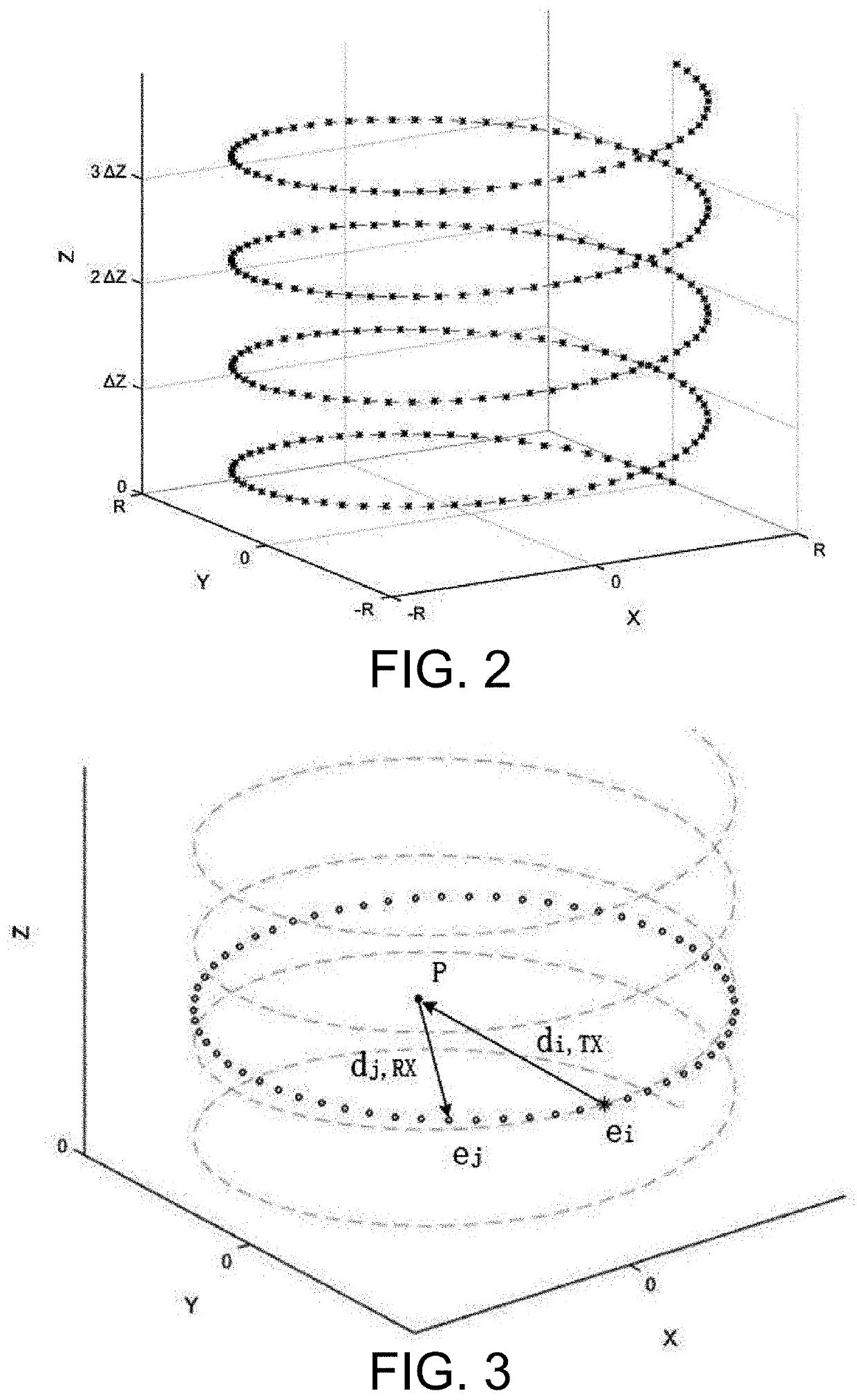
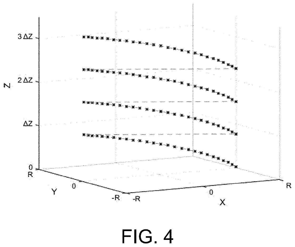
[0052]Uniformly distributed array elements on a ring detector are numbered from 1 to N, where N is the number of array elements in the detector. A period of time before starting the collection, the detector is drawn by a motor for uniform motion (the direction of motion is defined as parallel to the Z-axis direction of a spatial three-dimensional direct coordinate system), and the speed is denoted as S. When the collection is started, an array element 1 emits an ultrasonic wave, is received by all N array elements. After a time interval T, an array element 2emits an ultrasonic wave, and is received by all N array elements. Sequentially, after an array element N has emitted, it cycles back to the array element 1 to emit an ultrasonic wave until a scanning area covers an imaging object, and scanning is stopped.
[0053](2)...
embodiment 2
[0070]The three-dimensional ultrasound tomography method based on spiral scanning in the embodiment specifically includes the following steps.
[0071](1) Collecting data:
[0072]It is assumed that the central angle corresponding to the arc of a part of the ring detector is θ0, and the radius of the arc is R. Uniformly distributed array elements on the part of the ring detector are numbered from 1 to N, where N is the number of array elements in the detector. A period of time before starting the collection, the detector is drawn by a motor for uniform motion (the direction of motion is defined as parallel to the Z-axis direction of a spatial three-dimensional direct coordinate system), and the speed is denoted as S. When the collection is started, an array element 1 emits an ultrasonic wave, and is received by all N array elements. After a time interval T, an array element 2 emits an ultrasonic wave, and is received by all N array elements. Sequentially, after an array element N has emit...
embodiment 3
[0090]The three-dimensional ultrasound tomography method based on spiral scanning in the embodiment specifically includes the following steps.
[0091](1) Collecting data:
[0092]Uniformly distributed array elements on a ring detector are numbered from 1 to N, where N is the number of array elements in the detector. A period of time before starting the collection, the detector is drawn by a motor for uniform motion (the direction of motion is defined as parallel to the Z-axis direction of a spatial three-dimensional direct coordinate system), and the speed is denoted as S. After the collection is started, an array element 1 and an array element 2 emit ultrasonic waves at the same time, and is received by all N array elements. After a time interval T, an array element 3 and an array element 4 emit ultrasonic waves, and is received by all N array elements. Sequentially, after all emissions are completed, it cycles back to the array element 1 and the array element 2 to emit ultrasonic waves...
PUM
 Abstract
Description
Claims
Application Information
 - R&D
- Intellectual Property
- Life Sciences
- Materials
- Tech Scout
- Unparalleled Data Quality
- Higher Quality Content
- 60% Fewer Hallucinations
Browse by: Latest US Patents, China's latest patents, Technical Efficacy Thesaurus, Application Domain, Technology Topic, Popular Technical Reports.
© 2025 PatSnap. All rights reserved.Legal|Privacy policy|Modern Slavery Act Transparency Statement|Sitemap|About US| Contact US: help@patsnap.com