
Systems and methods for improving soft tissue contrast, multiscale modeling and spectral ct

Active Publication Date: 2022-08-11
ELUCID BIOIMAGING INC
View PDF13 Cites 5 Cited by
  • Summary
  • Abstract
  • Description
  • Claims
  • Application Information

AI Technical Summary

Benefits of technology

The invention is a computerized method for analyzing soft tissues by using different excitation sources, such as x-ray energy, radio frequency pulses, or ultrasound frequencies. The method involves obtaining multiple radiological images of the patient and selecting the best process to analyze the data based on the expected tissue type. The method can also involve segmenting the data to display the soft tissues and determining the tissue type in different regions of interest. The technical effects of the invention include improved soft tissue analysis and the ability to assess atherosclerotic plaque tissues in the vessel wall and perivascular space.

Problems solved by technology

Atherosclerosis can be life threatening, particularly in aging populations, but even among the relatively young.
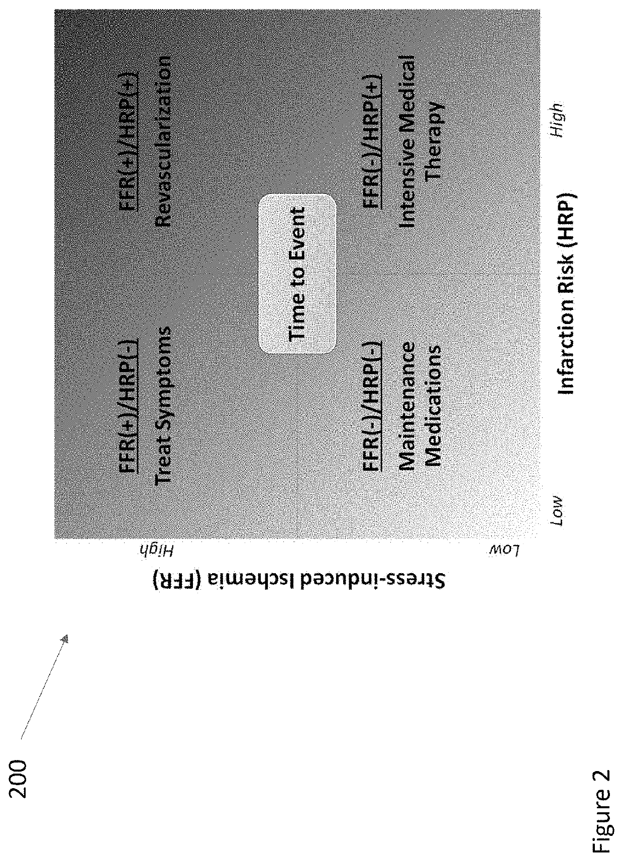
Current methods for diagnosing atherosclerosis, for example, the use of blood markers e.g., cholesterol levels) and / or determining the degree to which the lumen is narrowed (stenosis) are limited, and thus can result in suboptimal treatment decisions (e.g., to perform or not to perform surgeries, or prescribe intensive medical therapy).
For example, many vascular surgeries do not benefit the patient, some that need surgeries don't get them, and many could be effectively treated with drugs but may not be prescribed them.
Current tools can analyze a blood vessel lumen, but this can be insufficient for truly diagnosing atherosclerosis, as atherosclerosis is a disease of the vessel wall, rather than the blood or the channel through which it flows.
High rates of misclassified risk level, inability to assess likely response to drug therapy, and / or inability to measure response to drugs can occur.
With newer high-resolution imaging techniques, unaided, the radiologist can “drown” in data.
One difficulty with current imaging of atherosclerosis can include lack of robustness in the method used.
For example, current methods typically only provide a low level of contrast between blood vessel outer wall and perivascular tissues, thus making it difficult to distinguish between the two.
Vessel tapering, branching vessels, nearby tissues, etc. can also be problematic.
Another difficulty with current imaging of atherosclerosis can be due to a particular imaging device interrogating tissue using a limited excitation, and that despite the utility of multi-contrast MR on the one hand, or multi-energy CT on the other, the result can be a degree of non-specific response in the produced signal.

Method used


Image

  • Systems and methods for improving soft tissue contrast, multiscale modeling and spectral ct
  • Systems and methods for improving soft tissue contrast, multiscale modeling and spectral ct
  • Systems and methods for improving soft tissue contrast, multiscale modeling and spectral ct

Examples


Embodiment Construction

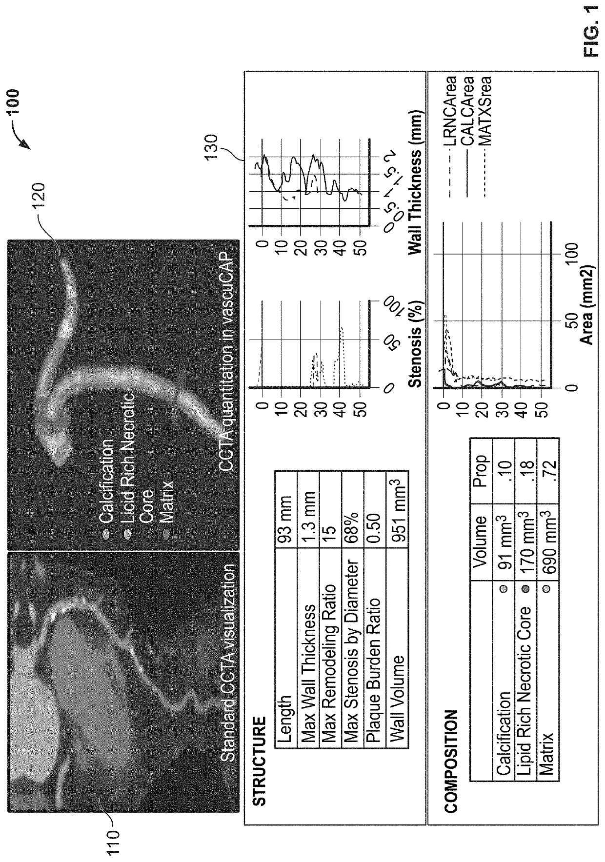
[0045]In general, the invention involves systems and methods for soft tissue contrast, characterizing tissue, classifying phenotype, stratifying risk, and / or performing multi-scale modeling aided by multiple energy or contrast excitation and evaluation. The invention can involve application of single vs. multi-phase image acquisitions and can include broad spectrum spectral CT to, for example, assess atherosclerotic plaque tissues in a vessel wall and / or perivascular space.

[0046]In general, the invention can involve exploiting differing responses by tissue to multi-energy or spectral image sets using a software approach. The invention can also include a hardware configuration that can further discriminate tissues in clinically relevant ranges. The spectral images obtained via the multi-energy levels and / or a broad spectrum can form the input for one or more algorithms that can improve tissue segmentation.

[0047]In some embodiments, the algorithm can include a exploiting a non-linear ...


PUM


Abstract

Systems and methods for improving soft tissue contrast, characterizing tissue, classifying phenotype, stratifying risk, and performing multi-scale modeling aided by multiple energy or contrast excitation and evaluation are provided. The systems and methods can include single and multi-phase acquisitions and broad and local spectrum imaging to assess atherosclerotic plaque tissues in the vessel wall and perivascular space.

Description

CROSS REFERENCE TO RELATED APPLICATIONS[0001]This application claims the benefit of and priority to U.S. provisional patent application No. 63 / 147,609, filed on Feb. 9, 2021, the entire contents of which are incorporated herein by reference in its entirety and owned by the assignee of the instant application.GOVERNMENT RIGHTS[0002]This work was supported in part by the National Heart, Lung, and Blood Institute, contract number HL 126224. The government may have certain rights in the invention.FIELD OF THE INVENTION[0003]The invention relates to computer-aided phenotyping (CAP) of disease which can include applying computerized image analysis and / or data fusion algorithms to patient data. In particular, the invention relates to quantitative imaging and analytics for elucidating the disease process of atherosclerosis, including techniques for improving soft tissue contrast, multi-scale modeling and / or spectral CT.BACKGROUND OF THE INVENTION[0004]Atherosclerosis can be life threatening...

Claims


Application Information

Patent Timeline
IPC IPC(8): G06T5/50A61B6/00G06T7/174G06T7/00
CPCG06T5/50A61B6/482A61B6/4241A61B6/5247A61B6/504A61B6/463G06T2207/20216G06T7/0012G06T2207/10081G06T2207/10088G06T2207/10132G06T2207/20224G06T7/174A61B6/5217A61B6/032A61B8/5261A61B8/481A61B6/4417A61B6/481A61B5/055G06T7/11
Inventor BUCKLER, ANDREW J.JI, CHANGGUOAYYAPILLAI, MURALI
Owner ELUCID BIOIMAGING INC
Learn More