
Enhanced Thermal Digital Subtraction Angiography (ETDSA)

a technology of thermal digital subtraction and angiography, which is applied in the field of medical imaging, can solve the problems of inability to use fluoroscopy and ct in early pregnancy, many temporary side effects, and inconvenient use, and achieves the effect of safe for patients and cheaper us

Active Publication Date: 2022-05-19
SULTAN HASHEM
View PDF3 Cites 0 Cited by
  • Summary
  • Abstract
  • Description
  • Claims
  • Application Information

AI Technical Summary

Benefits of technology

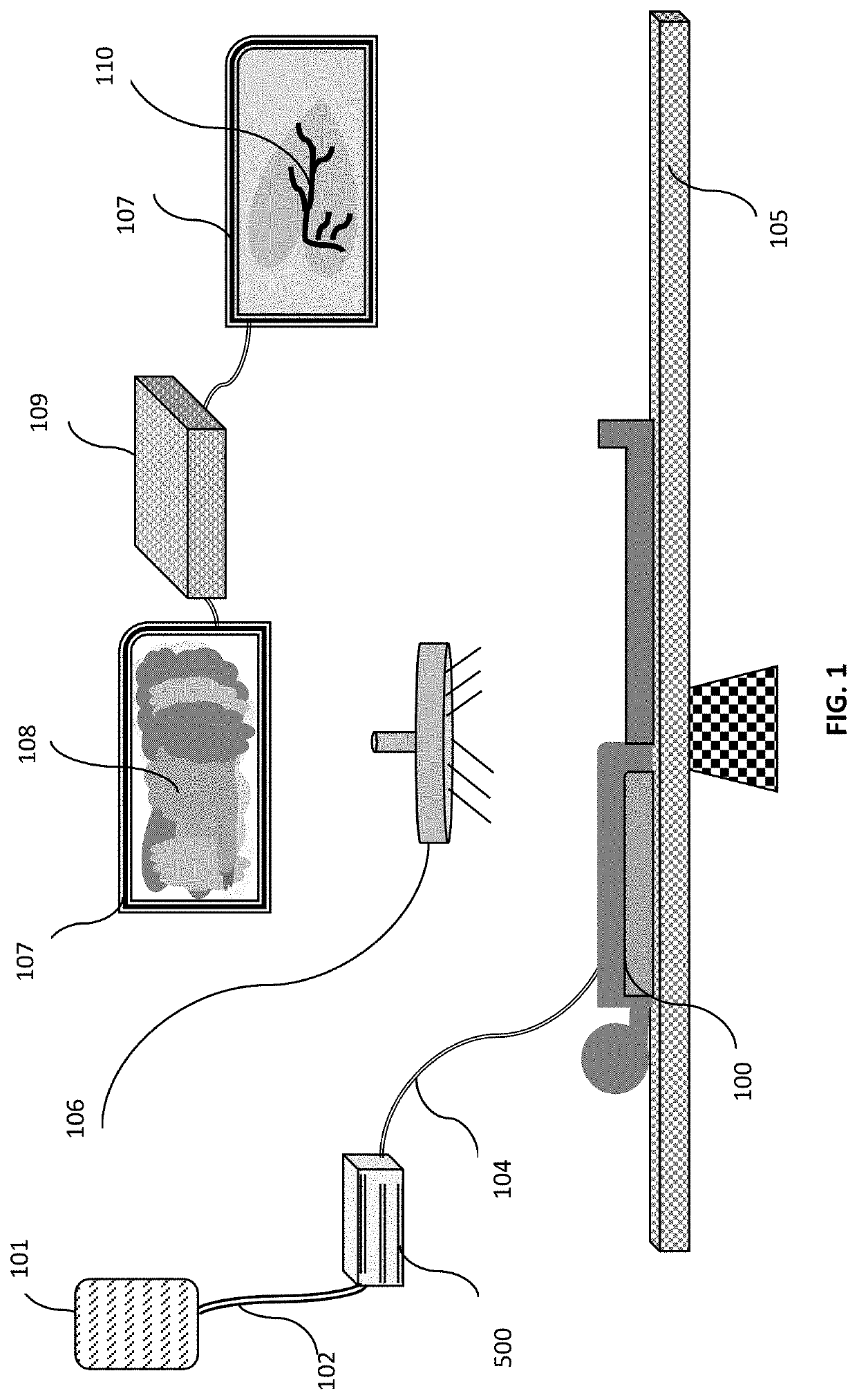
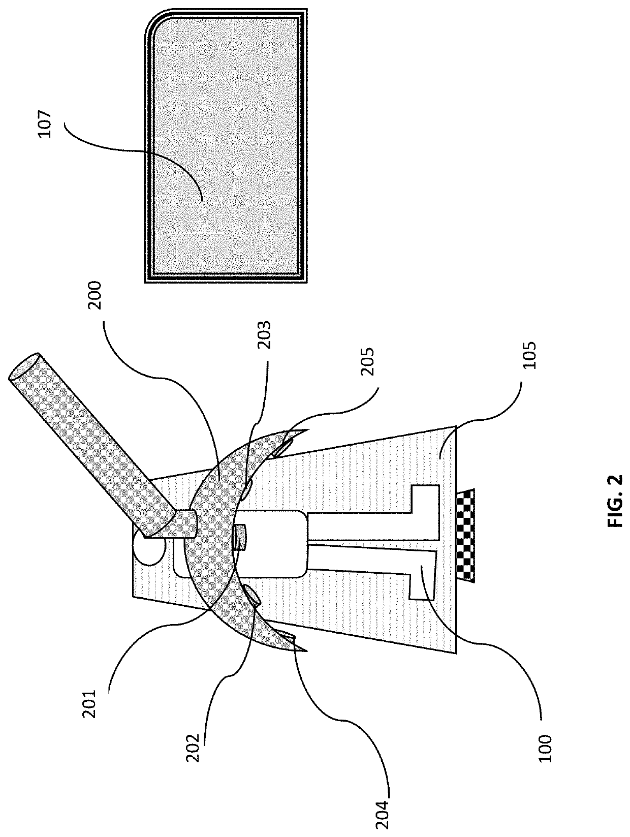
The current invention, ETDSA, is an improved technology for visualizing blood vessels that is safe, cost-effective, and can be used for both diagnosis and treatment. The technology uses a heat-conducting wire embedded in the catheter to improve thermal image quality. The invention also offers an advantage over X-ray and CT angiography for patients with chronic renal disease, allergic reactions to contrast agents, and pregnancy. It allows for emergency angiograms to be performed at the bedside without the need for arterial catheterization. The invention is portable, easy to use, and displays vascular images in color or 3D. It also measures the diameter, volume, and flow rate of specific vessels, as well as resistance and pressure differences.

Problems solved by technology

The images obtained before DSA was discovered were useful for determining anatomical position and variations, but unhelpful for visualizing blood vessels accurately.
Contrast material injected intravascular may cause nephrotoxicity, allergic reactions, and many temporary side effects.
Fluoroscopy and CT cannot be used in early pregnancy and can expose all patients to high radiation dose.
Thermal imaging's use in medicine had been very limited due to the fact that its images are hard to read due to the fact that IR images are inherently vague and unclear in nature.
However, its use in medical field did not match its use in other fields.
However, the image was limited in its clarity due to lack of subtraction of the images of the surrounding tissue.
Furthermore, this technique is limited to gastric bypass surgery when the blood is cooled to a very low degree to stop the heart from pumping during surgery.
Till today, there have been very slow progress in implementing and using thermal images in medical practice and very little progress had been made in this field despite the fact that IR camera was first invented in 1929.
This is due to the fact that its images are hard to read because IR images are inherently vague and unclear in nature.
The citations mentioned in this application are inadequate attempts to make a good use of this amazing technology.

Method used


Image

  • Enhanced Thermal Digital Subtraction Angiography (ETDSA)
  • Enhanced Thermal Digital Subtraction Angiography (ETDSA)
  • Enhanced Thermal Digital Subtraction Angiography (ETDSA)

Examples


Embodiment Construction

[0052]The current availability of the digital technology and science made it possible today to make further advance in the use of thermal images. As mentioned earlier, the application of thermal imaging in the field of medicine had been very limited mainly by its inherent nature of poor quality of images it produce, by the overlapping of images produced by the tissues surrounding the area of interest and by the difficulty in interpreting the images due to its complexity. Furthermore, the availability of high-quality imaging techniques such as MRI, CT, fluoroscopy, PET scan, ultrasound, made the interest in thermal imaging in medicine almost zero.

[0053]The current invention will put thermal imaging technology in its proper place in the medical field. This will give physicians more imaging options and provide additional advantages that other technology cannot offer. Example of such an additional advantage is that no other current technology can offer is arthrography and pulmonary angi...


PUM


Abstract

A method and a device for performing enhanced thermal digital subtraction angiography comprising infrared video camera, computer cable of subtracting all images produced by tissues having a temperature below a setup point, reconstructing and displaying 2D or 3D images of the received data, performing measurement of the vessels being displayed, conducting data analysis and mathematical calculation of hemodynamic parameters, storing and displaying the images produced. Said device is coupled to embodiments to warm the intravascular injectate, to warm the J-tip guide wire and to warm the intravascular catheter during angiography and during endovascular procedures.Furthermore, the device can be used to perform non-enhanced thermal digital subtraction imaging of part of interest and enhanced thermal digital subtraction imaging for joint, epidural and intrathecal space producing thermal arthrography, epidurography and myelography.

Description

BACKGROUND OF THE INVENTION1—Field of the Invention[0001]The present invention relates to medical imaging. Specifically, the current invention is related to image processing during angiography using enhanced warm fluids and digital subtraction of irrelevant tissues and organs.2—Description of Prior Art[0002]Angiography is a medical imaging technique used to visualize the inside of blood vessels with particular interest in the large and small arteries, veins and the heart chambers. This is traditionally done by injecting a radio-opaque contrast agent into the blood vessel. Different types of imaging had been in use; Fluoroscopy (Imaging can be done using X-ray based techniques), angiography, MRA (MRI angiography), radionuclide angiography (is an area of nuclear medicine which specializes in imaging to show the functionality of the heart. It involves use of radioactive agent, injected into a patient, and a gamma camera for acquisition.[0003]The angiogram technique was first developed ...

Claims


Application Information

Patent Timeline
Patent Type & Authority Applications(United States)
IPC IPC(8): G06T7/00G06T5/50A61B6/00
CPCG06T7/0012G06T5/50A61B6/504G06T2207/20224G06T2207/30101G06T2207/30024G06T2207/10048
Inventor SULTAN, HASHEM
Owner SULTAN HASHEM
Learn More